Tuesday, January 30, 2007

This is an 89 year old previous smoker with a probably stage 1b cancer:
No adenopathy.
She looks younger than her stated age but has emphysema on CXR:

We dont have PFT's but she has good exercise tolerance.

How would you proceed assuming the patient was willing to go with whatever the doctor recommends:
1. surgery (is age alone an absolute contraindication, even if she would otherwise be a surgical candidate)?
2. CT-guided bx (but what would you do with this info? it is obviously a cancer. Would you offer this 89 year old chemo?)
3. Do nothing.
4. Other.

Looking for all opinions - use anonymous or make up a name if you wish, but all comments encouraged.

Thursday, January 18, 2007

Solitary pulmonary nodule

I recently saw a 62-year-old woman with a past medical history of fibromyalgia who received a CT scan for chest pain, which picked up an incidental pulmonary nodule.
She is asymptomatic; no shortness of breath or cough. She is a 35 pack-year smoker and has not been able to quit. Her spirometry is normal.
A CT is shown here


What would you do?
1. refer to CTS for resection
2. Get a PET (and what would you do with that info?)
3. Follow radiographically, say, with a repeat CT in 3 months?

I'll tell you what I did after comments.
Feel free to enter your name as "anonymous".....

Monday, January 15, 2007

Follow up to vocal cord paralysis

A new question from anonymous regarding the case on vocal cord paralysis

Has anyone scoped his GI tract to R.O esophageal disease?

multiple pulmonary nodules in system lupus

Case from slkanowitz:

57 y.o. white female with SLE for 20+yrs presents with multiple small non calcified nodules on chest CT. CT was done for evaluation of asthma. Pt. symptomatic only with her usual migratory pleuritic pain associated with her lupus, and mild, intermittent asthmatic symptoms present since young adulthood. Asthma is treated prn with albuterol inhaler. Largest nodule is 6mm, most are peripheral and they are bilateral. Follow up chest CT scan 4 mos. later shows resolution of some of the nodules, the remaining ones show no growth. PFT are normal, O2 sat. normal. There has been no change in symptoms and no change in treatment for her lupus during that time. Other pertinent medical problems include allergic rhinitis, chronic sinusitis, S/P antral windows, and chronic bronchitis. Negative for pneumonia, SLE pneumonitis, RA, ANCA, anti-phospholipid AB. Recent echo WNL. No hx malignancy.
Hx significant for living in Ohio River Valley prior to 1973 and Southern Calif. during 1990, but no history unusual respiratory infections.

1998 chest xray normal. No xrays done until CT scan of chest and sinuses in 09/06.

SLE confirmed without evidence of other connective tissue disease. Pt. on Placquenil 200mg BID and Methotrexate 22.5 mg/week.


She did not pose a question but I assume it is what to do about the multiple nodules.
Since the nodules decreased in size over 4 months, this is likely inflammatory and a repeat CT in 6 months for that last nodule would be what I would do. I'm not sure what her smoking history is, but pretest prob for malignancy is low here. Any one else wish to comment?

Tuesday, January 09, 2007

Hypersensitivity Pneumonitis

Anonymous M.D. asks if anyone knows of alternative immunotherapy for the treatment of chronic hypersensitivity pneumonitis in a patient who is not responding to steroids. Her FVC has remained in the mid 50% range regardless of predxnisone does (range 0 to 40 mg). The HRCT shows fairly extensive ground glass to indicate an active alveolitis. Of note, this patient has an open lung bx that was consistent with the diagnosis of HP).

Tuesday, January 02, 2007

Follow-up to PAP case

I had posted a while ago on this patient with alveolar changes and a Bx consistent with PAP. We had also initiated a discussion on PAP and anti-GM-CSF antibodies.
I did send the Pt's blood to the Cleveland Clinic and her titers were 1:12,800 (1:400 or less is the usual normal level).
She is still asymptomatic and doing well.
Have you checked anti-GM-CSF ABs in your PAP Pts? Would you start treatment at this point (she is asymptomatic with normal PFTs) or just monitor?

Post-thoracotomy pain

One of my patients has had persistent post-thoracotomy pain following a RULobectomy in 9/05. She has been on gabapentin and narcotics; tried nerve root blocks (with short-lived results) and is not a drug-seeker. She has now undergone a dorsal rhizotomy (confirmed by path of the dorsal root ganglion) and after a 1-week improvement is now having even more pain.
What do you usually do for these cases of persistent post-thoracotomy pain that extend for months and years?

Wednesday, December 27, 2006

Portable oxygen concentrators

One of my COPD patients was asking me about purchasing a portable/travel concentrator such as this one.
Have you had much experience or feedback with portable oxygen concentrators?

Tuesday, December 19, 2006

Vocal cord paralysis

This 61 y/o man was sent to our office by ENT for w/up of a vocal cord paralysis. He has had subacute-onset hoarseness for the past 2-3 months and ENT found a unilateral R vocal cord paralysis. He did a CT (see the images below), which only showed some mediastinal and hilar (R>L) adenopathy much of which is calcified.
He has had a PET scan which showed some very faint (SUV<4) style="display: block; text-align: center;" alt="" src="http://photos1.blogger.com/x/blogger/7040/842/320/933316/Vocal.jpg" border="0">

Friday, December 15, 2006

RLS

Have you had a lot of experience with Requip (ropinirole) for restless leg syndrome?
If not, what is your preferred therapy fro RLS?

Monday, December 11, 2006

Differential and w/w

43 year old presented with 6 weeks of progressive dyspnea associated with URI-type symptoms. Had recieved several courses of antibiotics as an outpatient. No fever/chills. + non-productive cough. No associated s/s. Non-smoker. No occupational exposure. On presentation, he was hypotensive, but alert and oriented. His was quite comfortatble despite profound hypoxia (initial oxygen saturation of 62% on room air). Physical exam has some scattered rales. WBC is 4, without a shift. Blood count and platelets are normal. He is hypoxic, requiring up to 100% supplemental oxygen by non-rebreather to maintain a pO2 in the 60's. His ventilation is adequate, with a pCO2 of 42 and a pH of 7.39.

A CT scan shows:


Saturday, December 09, 2006

Follow-up to Pulmonary Nodules

This is the 51 y/o woman with COPD and pulmonary nodules.
We had the same concerns and DDx as proposed in the discussion. I did a bronchoscopy and we got good samples consistent with RB-ILD.
She was counselled again regarding TOB cessation and agreed to try varenicline. She will follow-up in a few weeks and we will monitor her CxR and PFTs.

Tuesday, December 05, 2006

Pulmonary nodules

This is a 51 y/o woman followed in our office for moderate COPD. She continues to smoke and has had worsening dyspnea and weight loss for a couple months. Her CxR and CT scan, which in the past showed hyperinflation and nothing else, now have these pulmonary nodules.
What is your DDx and wht would you do next? Posted by Picasa

Thursday, November 30, 2006

Airway remodelling

I have posted a question on this a while ago. I see a fair number (just saw one today) of older patients (usually women) who have never smoked, have normal gas exchange and chest radiography but who present with moderately severe degrees of fixed obstructive lung disease. They never have any history of asthma, cough or wheezing and their only complaint is DOE.
Usually there is some small-airway obstruction on the expiratory images of a HRCT but no other structural abnormalities.
Have you seen much of that? How do you label these patients? Unsuspected asthma with remodelling?

Monday, November 27, 2006

SOB

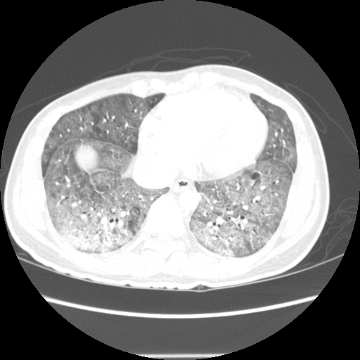
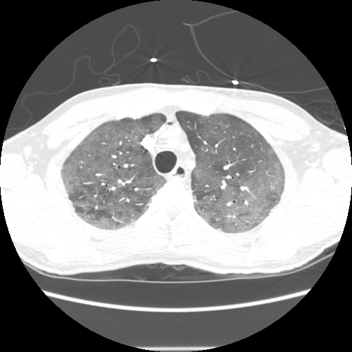
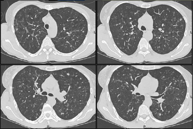
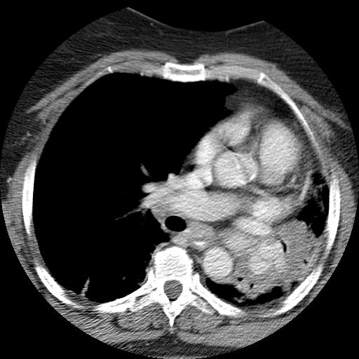
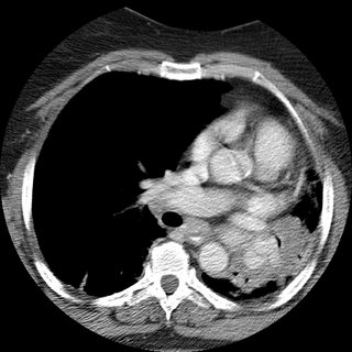
62 woman c/o 2 weeks ago started to have cough with greenish sputum. She presented to the ER where workup included a CT (below).
Pt states that was diagnosed with asthma in 2002; at that time she had a CXR that showed scar tissue in the left upper lobe secondary to a previous pneumonia.
She noticed that over the last year, she has being unable to lay down over her right side because she would feel short of breath, so that she always sleep over her left side. Denies frequent cough, sputum production, hemoptysis, weight loss, loss of appetite, chronic chest pain.
SOCIAL HISTORY:
pt has 40 pack year history of smoking, quit in 1998. She works as hair stylist and is exposed to fumes and chemicals. Denies exposure to TB. No recent travels.
PHYSICAL EXAM:
BP 139/83, HR 86x', RR 16x', O2Sat 93% on RA, W 212 pounds.
Patient is awake, alert, obese, oriented x3, no acute distress.
Neck, no lymphadenomegaly. Chest, no use of accesory muscles, no retractions, flat to percussion on the left, no prolonged expiration, absent breath sounds in the left, no wheezes, no crackles. CV: regular rhythm, no S3 or S4, no murmurs. No rubs.

Spirometry showed severe obstruction.

(quick) DDx and how would you proceed?

Friday, November 24, 2006

Follow-up to dyspnea

This is the recent post of the 47 y/o woman with dyspnea. JJ and Krayem suggested HP and on the TBBx we had loose granulomata with lots of lymphocytes and negative organisms on stains and Cxs. The pathologist felt quite confident calling it as HP. Unfortunately I don't know what is the culprit exposure yet...

Tuesday, November 21, 2006

CXR followup after resection

A curious reader from Maine would like to know:
How frequently and for how long, do you follow xray/CT's after the surgical resection of a stage 2 lung cancer.

BCG and latent tb

A patient had a previous negative PPD as a requirement for work. She then went oversees for a time and receieved the BCG vaccine. She is now back in this country and as part of work requirements gets another PPD which is now positive. She is asymptomatic and the chest xray is negative.
How would you handle this?

Dyspnea


I have just seen this 47 y/o woman for new onset dyspnea. She has had malaise, non-productive cough and DOE for the past 6 weeks. Occasional "low-grade" temps but no fevers.
No TOB Hx. She works at a auto air-bag manufacturer and part-time at a Wal-Mart. No other exposures. Exam is pretty unremarkable. PFTs revealed a mild restrictive deffect with low DLCo.
Any suggestions? (BTW, ignore the small apical PTx, that was from vigorous bronching). Posted by Picasa

Wednesday, November 15, 2006

Dyspnea


I just saw this 70 y/o woman in consult today. Her only complaint is dyspnea on exertion: she gets tired after walking more than a mile. No TOB, no Hx of lung Dz.
She had 3-4 episodes of pneumonia within 2 years > 5years ago.
No childhood illnesses.
No chronic cough. Physical exam is unremarkable. PFTs showed a moderate mixed defect.
She has the bronchiectatic/focal cystic changes above and the rest of the CT was normal.
How would you work up her focal bronchiectasis and do think those small areas are sufficient explanation to her dyspnea.