
Friday, June 26, 2009
Monday, August 25, 2008
Intravenous haldol in the ICU
This has come up on our recent P&T meeting. We use a fair amount of IV haldol in the ICU even though haldol is not FDA-approved for IV use (that has never bothered us before). However, the FDA has now posted a new re-warning (to help us, I am sure):
Although injectable haloperidol is approved by the FDA only for intramuscular injection, there is considerable evidence from the medical literature that intravenous administration of haloperidol is a relatively common “off-label” clinical practice, primarily for treatment of severe agitation in intensive care units. Due to a number of case reports of sudden death, TdP and QT prolongation in patients treated with haloperidol (especially when the drug is given intravenously or at doses higher than recommended), the sponsor has updated the labeling for haloperidol. The updated WARNINGS note that:
* Higher doses and intravenous administration of haloperidol appear to be associated with a higher risk of QT prolongation and TdP.
* Although cases of sudden death, TdP and QT prolongation have been reported even in the absence of predisposing factors, particular caution is advised in treating patients using any formulation of haloperidol who:
- have other QT-prolonging conditions, including electrolyte imbalance (particularly hypokalemia and hypomagnesemia)
- have underlying cardiac abnormalities, hypothyroidism, or familial long QT syndrome
- or are taking drugs known to prolong the QT interval.
* Because of this risk of TdP and QT prolongation, ECG monitoring is recommended if haloperidol is given intravenously.
* Haloperidol is not approved for intravenous administration.
Pharmacy has been concerned on how to include this in policy. Do you use much IV haldol?
Wednesday, June 04, 2008
Inhaled LABA's
Here's a link to a new meta-analysis (Annals of Internal Medicine) of LABA's + ICS vs. ICS alone. It's taken from the GSK database, and finds the significant increase risk to the addition of Salmeterol to ICS with regards to mortality, asthma hospitalization, or asthma exacerbation.
Contradicts the findings from the Salpeter meta-analysis and from SMART (which I did not think were very compelling to begin with). So, any thoughts? Does this affect management of patients with asthma?
Here's the link to the paper and it's editorial:
http://www.annals.org/cgi/content/abstract/0000605-200807010-00229v1?papetoc
http://www.annals.org/cgi/content/full/0000605-200807010-00230v1?papetoc
Wednesday, May 28, 2008
Adenopathy
Patient is a 68 year old African American woman with a history of non-Hodgkin's lymphoma treated with chemotherapy and radiotherapy in 1980.
Symptoms now: she has type-B symptoms only - sweats at night, lost 14 pounds in the last 2 months, 7 pounds in the last 1 month. Low appetite also. There are no respiratory symptoms at all.
She also has burning feeling in the feet
She has right hilar and subcarinal adenopathy; she also has gastrohepatic and periportal adenopathy and slightly more prominent intrahepatic biliary tree dilatation with slightly more dilatation of the previously dilated common bile duct.
A surgical excision biopsy of an axillary node showed non-caseating granulomatous inflammation with no lymphoma at all.
Again her symptoms are as above - some B symptoms and a likely peripheral neuropathy (I think).
How would you approach this patient next?
Thursday, May 22, 2008
Update on cavitary lesion
Well, I thought this patient would have active TB. 3 AFB's were negative. Here is a CT we then got:


We bronched her and all AFB's are still negative, 6 days later. The TBBX was also negative:
minimal inflammatory cells. Bronchial mucosa and submucosal glands are unremarkable. Alveoli show few pigment laden macrophages. No evidence of granulomas or viral inclusions is seen. AFB stain is ordered, see addendum.
She is currently taking 4 drug TB therapy (after bronch)
What would you do now??
Thursday, May 15, 2008
What do you see?
The patient is a 59-year-old female with history of hepatitis C, currently on dual therapy with pegylated interferon and ribavirin for last 2 months presented with complaint of cough.
PAST MEDICAL HISTORY:
1. Hepatitis C, liver cirrhosis.
2. Childhood asthma.
3. History of positive PPD more than 10 years ago, which was positive, at
that time with negative chest x-ray.

PAST MEDICAL HISTORY:
1. Hepatitis C, liver cirrhosis.
2. Childhood asthma.
3. History of positive PPD more than 10 years ago, which was positive, at
that time with negative chest x-ray.

Monday, May 12, 2008
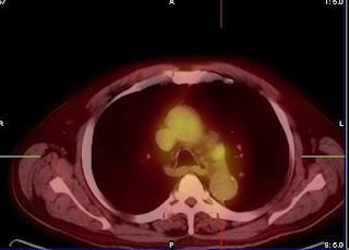
stage iiia?
Would this PET scan be enough to convince you that this is IIIa or would you have the surgeon go after that node for staging purposes?
A corollary: if that node is negative, would he be a surgical candidate (assuming lung function etc. not issue)

A corollary: if that node is negative, would he be a surgical candidate (assuming lung function etc. not issue)


Saturday, April 05, 2008
post thoracotomy pain
One more post today. How do you best deal with post-thoracotomy pain. I admit I see it fairly often as well.
Patient writes in a recent comment
I have acute persistent pain in my right breast and along the six inch incision at the base of my breast. I have tried gabapentin, Lyrica, and Topimate. I am on my third round of corticosteroid injections into the intercostals. I am reluctant to use narcotics, but if the steroids do not work, I am at a loss. I cannot move my right arm without pain. Any suggestions would be greatly appreciated.
ABPA without steroids
A reader writes:
I am 48 years old female, and lived with asthma for the last 25 years. I am not under great control. I have had blood work done recently, and found out I have severe allergies. I have an IgE of 1700, my aspergillus allergen is 2.05 ku/l. I recently had a chest ct which showed nonspecific infiltrates with ground glass appearance. A sinus ct showed acute maxillary sinusitis. my physician says she is not sure of a diagnosis of ABPA, and is hesitant to treat me with steroids. In the past year I have been diagnosed with avascular necrosis of both hips, and my right knee as well as severe cataracts in both eyes. I was told probably from the use of steroids to treat my asthma. My concern is I never feel great, and why is this so hard to diagnose, and if I have ABPA what will ahappen if it goes untreated.
What types of steroid-sparing therapies do you use for ABPA (or asthma)?
Thursday, March 20, 2008
Acute ILD
This 77 year old man has had a bit of honeycombing at the bases since at least 2004. Radiographically c/w UIP. Biopsies not done and he has been stable for years (and FVC normal at 84% predicted).
In February he developed worsenign cough and SOB. He was admitted and was hypoxic with the CT shown below. A cath was done showing normal wedge, normal CO/CI.
A BAL showed only 184 cells half of which were macrophages. A TBBX was not done because he's on coumadin for afib and we didn't think it would add much to determining the diagnosis.
SH, desk job all his life. In January, routine maintenance change of the humidifier pipes in his furnace. This may be a red herring but throwing it out there. No one else in household sick.

Was going to send him for open lung/VATs, but what do you think are possible etiologies?
I also repeated autoimmune chemistries that had been negative in 2004.
Wednesday, March 19, 2008
lesion in a 45 year old woman
A 45 y/o woman presented with atypical chest pain that led to a ct showing the following:

Although a benign lesion/resolving pneumonia were possibilities, I was concerned about bronchoalveolar carcinoma in this young non-smoker. A bronch was done - BAL cytology and tbbx were negative. We were in the right area (posterior segment RUL) and Fluoro did support this, but the biopsies came back as unremarkable pulmonary parenchyma.
What would you do next? Some options would be follow with serial CT's - q 3 months, open lung, repeat bronch, or another course.
Wednesday, February 20, 2008
What's your line?
A resident put in a left subclavian line and maybe went a little too far. An xray shows that it went into the right atrium and into the IVC and up to the IVC filter. When it was removed (with help from vascular), the filter had migrated up 2 vertebrae... Click image to enlarge. It is a bit dark.


Friday, February 15, 2008
Fibrosis
A submission from a reader: "34 year old, non-smoking African American female presenting with a recurring (for three years now), chronic (lasts for several weeks up to a few months) dry, non productive cough. No other symptoms (though the attached CT report says wheezing). No response to a variety of drug treatments over the years, including steroid shot, Levaquin, OTC Mucinex, OTC Claritin, Tussionex, Nasacort, OTC Prilosec, 6-day regimen of Metrol (4mg), Advair and Albuterol. First pulmonologist (who performed no diagnostic tests) diagnosed asthma and allergies. Nothing indicated on chest x-ray. Internal medicine doctor ordered CT scan (report attached). Second pulmonologist said subpleural cysts exist, which he indicated exist with UIP patients, but he said this was not UIP. Also said it's not tuberculosis or sarcoidosis. Prescribed 20 mg of Prednisone twice a day to get rid of cough, said we will proceed once cough is gone. Said a lung biopsy or thoracic biopsy may be in order.
CT reading:
Helical CT images through the chest were obtained following
intravenous contrast administration.
Within both lungs and involving both the upper and lower lung zones,
there are areas of subpleural macrocystic change with some
associated subpleural interstitial thickening. There are no
particular areas of ground-glass opacity associated with these
findings. No significant nodularity is seen. The central and midlung
zones are spared. No pleural fluid or lymphadenopathy is seen. These
findings can be seen in patients with usual interstitial pneumonia
(UIP) although this would be somewhat atypical in a patient of this
age group. Does the patient have a smoking history? Other
interstitial lung disease in its early stages may give a similar
appearance.
Wednesday, December 19, 2007
41 year old with HIV and fever
This is a 41 year old man with AIDS, CD4 below 10, who presented with shortness of breath and fever. He was admitted to a general ward but trasnferred to the unit a few days later for tachypnea. On the general floor, his vitals were normal except for some tachycardia. He was 97% on 2 l NC. CXR:

a full workup was done to look for infectious source.
sputums: AFB negative x 3, fungal negative. PCP negative (no PCR, no bacteria
CSF - negative for infection.
What kinds of things could be causing these findings and what would you do?

a full workup was done to look for infectious source.
sputums: AFB negative x 3, fungal negative. PCP negative (no PCR, no bacteria
CSF - negative for infection.
What kinds of things could be causing these findings and what would you do?
Friday, November 30, 2007
Lymphocytic effusion
Since another question from a doc is related to TB, I'll post that here as well:
55 y old gentleman presented with few weeks history of progressive dyspne and right sided pleuritic chest pain, with history of contact with a case of pulmonary TB, no symptoms of toxemia, clinically the patient got signs of right sided pleural effusion which was aspirated and shown to be lymphocytic exudate,because the patient also presented with hoaseness of voice CT chest was done showed no lung masses or lymphadenopathy, BAL showed no malignant cells, PPD test was highly positive, laryngeal examination showed cordal polyp. culturing the fluid and sputum for TB was negativethe patient was started on antituberculous ttt , 1st 2 months quadrible therpy and then dual therapy and the patient still have re accumulating effusion? any suggestions?
55 y old gentleman presented with few weeks history of progressive dyspne and right sided pleuritic chest pain, with history of contact with a case of pulmonary TB, no symptoms of toxemia, clinically the patient got signs of right sided pleural effusion which was aspirated and shown to be lymphocytic exudate,because the patient also presented with hoaseness of voice CT chest was done showed no lung masses or lymphadenopathy, BAL showed no malignant cells, PPD test was highly positive, laryngeal examination showed cordal polyp. culturing the fluid and sputum for TB was negativethe patient was started on antituberculous ttt , 1st 2 months quadrible therpy and then dual therapy and the patient still have re accumulating effusion? any suggestions?
TB treatment and a followup BAL
A physcian from Florida recently asked how one should approach the following. A younger man from a TB-endemic area with cavitary upper lobe lesions. He is not productive of sputum. Obviously, the physician elected to treat empirically for TB. In terms of getting sensitivities, a BAL should be done, but his question was, how long after initiation of 4-drug therapy would the BAL give a false positive. By false positive, I guess you could view that as as either afb negative, or culture negative (if the former represents dead TB bugs).
He was considering waiting 2 weeks to help decrease the risk to those in the bronchoscopy suite.
What do you think?
He was considering waiting 2 weeks to help decrease the risk to those in the bronchoscopy suite.
What do you think?
Monday, November 12, 2007
need for lymph node biopsy?
Question submitted by anonymous:
24 year old Asian female presented with chronic productive cough of green/yellow sputum for the last year. Travelled to Malaysia and Pakistan in the last year. Some mild episodes of haemoptysis. CXR when the patient initially presented was NAD. Bloods all normal, barring a bilirubin of 16.
A CT a year after initial presentation showed right upper lobe collapse with a 2cm mass. Left upper lobe bronchiectasis. Also widespread mediastinal adenopathy.
Sputum cultures negative. Bronchoscopy showed a sputum plug sent for MC+S - negative. Nil else on bronchoscopy.
Why is there mediastinal adenopathy? Should a biopsy be performed in order to aid diagnosis?
Thursday, October 25, 2007
Lung cancer letter
Very interesting letter to the editor on lung cancer, that puts things into perspective and that is often overlooked. Written by someone who also has left many comments on this site, so check it out.
Tuesday, October 23, 2007
IPF treatment - worse than the "cure"
Here's another anecdote on a patient started on prednisone/azathioprine/NAC for IPF. He was started on this for a variety of reasons, but one was that the biopsy had a bit more inflammatory changes even though there was ample fibroblastic foci and heterogeneity, so I thought a 3-6 month trial would be reasonable. By the time his imuran was up to 75 mg, I saw him. A repeat spiro was unchanged (FVC 43% predicted) but his DLCO went from 35% to 45% so I continued the meds. However he had mouth pain and the tongue showed possible thrush so I kept the Imuran at 75, gave some nystatin swish and sent him out to be followed up in 2 months and repeat the HRCT with the sprio/DLCO. However, his mouth pain did not go away and worsened, and he started developing malaise and a fever. I saw him in clinic that day. He looked fairly sick but vitals ok. He had a soft palate lesion. I got derm to KOH it and there are some non-budding hyphae so he's to get clotrimazole . Then almost as an afterthought I added on amylase and lipase to the repeat LFT's and low and behold the lipase is 800.
Of note his WBC was 13 and now down to 6....
This side effect is likely going to be self-limiting as he stays off the imuran. As I tell him to go light on PO intake, I am adding a creatinine to make sure he is not volume depleted (he's an outpatient).
Of note his WBC was 13 and now down to 6....
This side effect is likely going to be self-limiting as he stays off the imuran. As I tell him to go light on PO intake, I am adding a creatinine to make sure he is not volume depleted (he's an outpatient).
Monday, October 15, 2007
Precedex?
Redneck Crit Care (nice name) submitted this question:
One of our CT surgeons has been using Precedex for postop sedation for vent patients with good success.
www.ptjournal.com/ptjournal/fulltext/30/3/PTJ3003158.pdf
according to information in that article, it appears to be a very attractive option. It is a short-acting alfa2 agonist and you do not have to discontinue this before, during or after extubation because it does not cause respiratory depression. Are many intensivists already using this in medical ICU?
Subscribe to:
Posts (Atom)