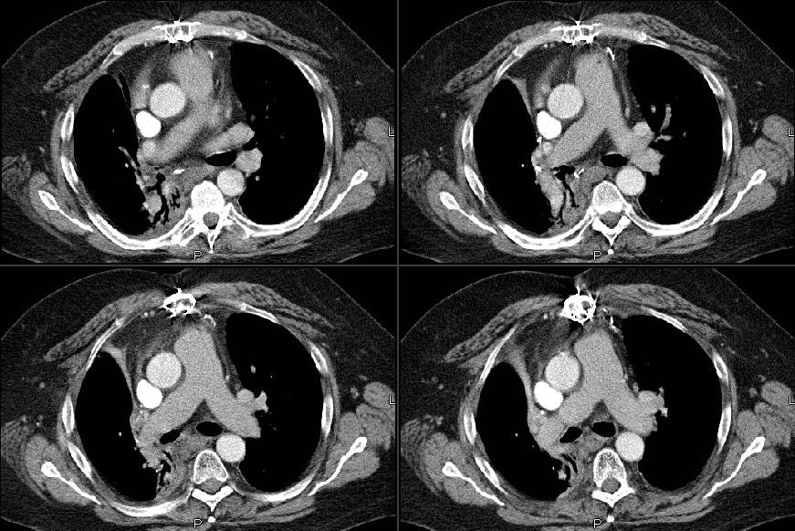
He came back to his PCP this past month with a new cough and R chest tightness. His CT showed some loss of volume and more soft tissue swelling in the hilar area (see below):

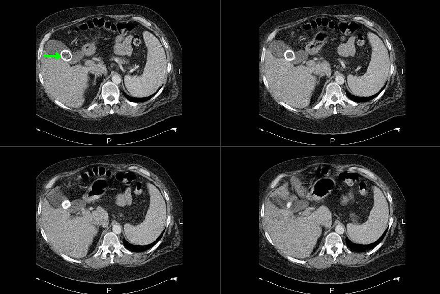
Incidentally he had this huge gallstone (cool pic):
I bronch'ed him and he had narrowing and scarring of the residual RML bronchus with normal RLL and a healthy looking RUL stump. EndoBBx, brushings and lavages were remarkable for radiation changes and atypia but no neoplasm. Would you do a PET, a more invasive sampling (e.g. Mediastinoscopy) or just follow him after some ABTx since he has already had maximal therapy to the area and would have limited options with recurrent Ca?
No comments:
Post a Comment