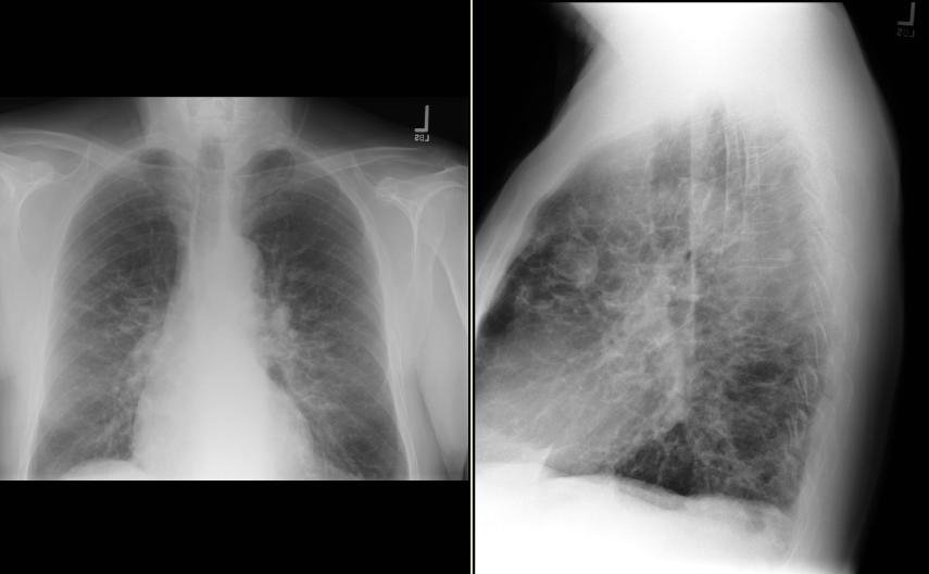
The xray is hard to read. Am I reading it correctly if I say there appears to be thickened lines, in a reticular pattern (seen best on the lateral) I don't want to go through the whole ddx if lower lobe ILD if the above is wrong. On the PA there appears to be some b/l haziness but I can't zoom in to see if there is an alveolar filling process or if this is all just interstitial dease in a "butterfly" pattern
It looks like your radiology dept has the same problem as ours... cuts off the costophrenic angles bilaterally. In addition to Jennings' comments, I would mention that the hilae look full with the left being more prominent than the right. This may explain the "nodule" visualized in the retrosternal area.
The xray is hard to read. Am I reading it correctly if I say there appears to be thickened lines, in a reticular pattern (seen best on the lateral) I don't want to go through the whole ddx if lower lobe ILD if the above is wrong. On the PA there appears to be some b/l haziness but I can't zoom in to see if there is an alveolar filling process or if this is all just interstitial dease in a "butterfly" pattern
ReplyDeleteThose are good comments, check out the lateral particularly the retro-sternal area.
ReplyDeleteIs there a rounded opacity/mass just anterior to the cardiac border?
ReplyDeleteIt looks like your radiology dept has the same problem as ours... cuts off the costophrenic angles bilaterally.
ReplyDeleteIn addition to Jennings' comments, I would mention that the hilae look full with the left being more prominent than the right. This may explain the "nodule" visualized in the retrosternal area.