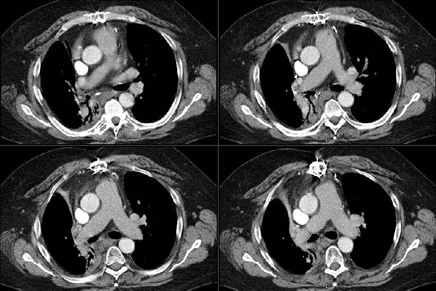
He came back to his PCP this past month with a new cough and R chest tightness. His CT showed some loss of volume and more soft tissue swelling in the hilar area (see below):

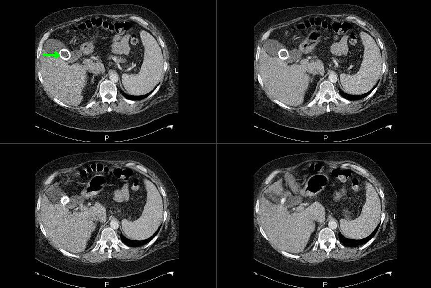
Incidentally he had this huge gallstone (cool pic):
I bronch'ed him and he had narrowing and scarring of the residual RML bronchus with normal RLL and a healthy looking RUL stump. EndoBBx, brushings and lavages were remarkable for radiation changes and atypia but no neoplasm. Would you do a PET, a more invasive sampling (e.g. Mediastinoscopy) or just follow him after some ABTx since he has already had maximal therapy to the area and would have limited options with recurrent Ca?
0 comments - CLICK HERE to read & add your own!:
Post a Commenttest post a comment