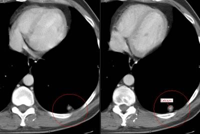
This is a 44 y/o man who works here in the hospital. He is a ctually a nice guy so I'm trying not to find anything too interesting on his case.
He is very healthy and smoked a pack-a-day for ten years but quit in 1988. He had travelled to the gul area and came back with a severe gastroenteritis. It seems that it was so bad the primary team got a CT of his abdomen. He did OK but the lower cuts of the lung revealed a ~1-cm partially calcified LLL nodule. He is completely asymptomatic from a respiratory perspective. His PFTs are completely normal. We got a dedicated CT of the chest and found the following nodules: (a 9-mm RUL a ~5-mm L apical and the same LLL).
The RUL has a little bit of excentric calcium as does the LLL one. Would you PET, biopsy, watch or else?
Sunday, July 30, 2006
Subscribe to:
Post Comments (Atom)


1 comments - CLICK HERE to read & add your own!:
The size of the other nodules means you'll just ignore a negative PET anyway. The other has some calcium but the way it is distributed within the nodule doesn't rule out Ca. I assume there are no nodes. I would repeat CT in 3 months and just follow if there is no chaneg in these 3 nodules. If there is increase, would PET at that point. Extra info at 3 months would be whether all 3 are growing or just the LLL nodule.
Post a Commenttest post a comment