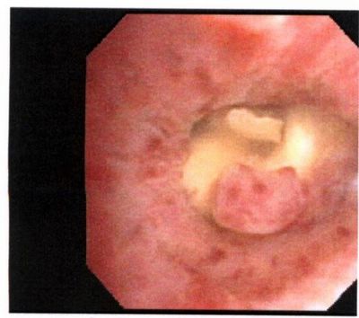
My partner bronch'ed this pleasant 78 y/o woman with no TOB Hx who had developed a persistent RML collapse. The first picture was taken while instilling NSS to keep the airway open.
The lesion was hard to biopsy and there was purulent mucus behind it.
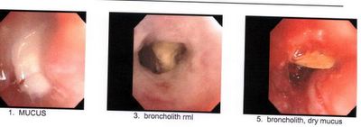
The second set of pictures was a re-bronch 2-3 days later after he tried to remove as much as possible with the forceps.
Any suggestion, comments?

3 comments - CLICK HERE to read & add your own!:
This nodule look like smooth surface which appears benign. Assuming no other symptons, is perhaps a benign polyp.
Biopsy results?
carcinoid?
It is actually a broncholith: a calcified limphnode eroding through the wall and causing RML collaps.
Post a Commenttest post a comment