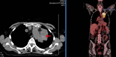
This is an 82 y/o woman with no smoking history but decades of second-hand smoke (her husband died of lung dz). She had a dry cough and a CT scan was done. She has the large LUL mass seen on the CT cut (on an outside CT with contrast, the mass seems to invade the PA). I did a bronch and the mass is a non-small cell Ca. Unfortunately, the hilar TBNA were non-diagnostic. Her PET scan shows marked uptake by the mas, ipsi- and contralateral hilar nodes and pre-tracheal nodes.
With the PA involvement (on CT) and the PET results would you be satisfied in calling it a IIIB or would you do a mediastinoscopy to confirm either/both with tissue?

3 comments - CLICK HERE to read & add your own!:
Perfect question for a multidisciplinary oncology conference. There are two potential lesions making it 3B--the PA invasion and the contralateral node. So, I'd say it depends on how clearly it's invading the PA---is it obviously crossing a tissue plane, or is it possible abutting, but not "invading"...
We are discussing all of our patients in multidisciplinary meetings once a week, this case would be discussed as well.
"Seems" to invade the PA: so I wouldn't personnally call it a IIIb on this finding unless invasion is obvious.
Ipsi/controlateral nodes: hard to say about the node activity level on this frontal slice. Anyway, PET-CT is especially interesting for its negative predictive value in nodes involvment: no uptake, more likely no invasion.
I would never claim for an abstention in front of a hot node, especially in some areas of the states where anthracosis and other inflammatory diseases are commun reasons of false positive cases. Specificity is higher than CT alone, but still not 100%.I would recommend a mediastinoscopy.
PET-CT isn t there to falsely waste surgery indications.
Should this patient be classified as a IIb after surgery, I would recommend a pet-CT in the follow up, and see how those contro-lateral nodes look like.
ps: why no contrast in this pet-ct?
Thank you for the commments. Our radiologists will usually do the combined PET-CT portion without IV contrast. Since she had just had a previous CT with IV dye we felt she did not need the other dye load.
We did discuss a med with the patient but she was very concerned with any surgery and would not want a lobectomy. She chose to go for multimodality Tx instead.
Post a Commenttest post a comment