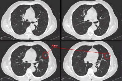
I had been following a 79 y/o man with small non-calcified nodules with serial CT scans. He is a lifelong non-smoker and one of his noudles was actually calcified and he had a calcified small hilar node. His nodules had been stable for about a year and with the lack of smoking Hx and other finding I was quite confident these findings were benign.
He developed a new URI and through his PCP, had a new CXR followed by a CT scan (see below). The CT revealed a small new left nodule. All the other previous findings had remained unchanged.
He then had a PET-CT: the nodule, and actually all the nodules as well, was PET-negative. However, the left hilum (not much there on CT was positive with an SUV of 2.54 and there was a positive subcarinal node with an average SUV of 2.2.
Are you very impressed by those numbers or would you write it off as inflammation and do a CT follow-up?
Monday, March 06, 2006
Subscribe to:
Post Comments (Atom)

2 comments - CLICK HERE to read & add your own!:
Yes with that history I would be quite comfortable ascribing the PET results to an inflammatory process. I would repeat the CT serially (next one in 3 mos) and follow from there.
Let me preface this by saying that I am *not* a doctor, nor am I even very experienced as a PET technologist, but what about doing a follow up PET exam in a few months, to be sure that it really is just inflammatory? The only reason I can think of for why you wouldn't is if there would be a problem with insurance covering it, but nothing like that is coming to mind.
Post a Commenttest post a comment