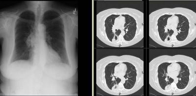
I usually don't like those boards-type question that ask "what would you do next?" and only let you choose one option since we often do more than one thing in real life.
Having said that, I am curious as to what you would do next in this case. This is kind of a VIP patient so she was referred to a couple different specialists at the same time.
She is in her early 70s, quit smoking in 1972, near normal PFTs (FEV1~70%, normal DLCO) and has an abnormal CxR. It started as an URI and then she developed a "deeper" cough with purulent sputum production and some pleuritic CP.
The abnormal CxR (and some CT cuts) are shown below.
She had no adenopathy on the CT.
What would be your first step?
Tuesday, October 17, 2006
Subscribe to:
Post Comments (Atom)

7 comments - CLICK HERE to read & add your own!:
I cant tell if thats a cavitary mass or airspace disease with cavitation, in the posterior segment of the right lower lobe. I would start with a bronch with BAL and TBBx for infections (including AFB, fungal bacterial) and Tbbx and send the BAL for cytology and the biopsy for path. A PET should probably be deferred at this point given that infection is also high in the differential still
How about some antibiotics and a repeat CXR?
I'd treat with antibiotics and repeat the CT in 6 weeks. If that consolidation is still there, I'd proceed with a diagnostic bronchoscopy.
Do you suggest bronchoscopy?
I had initially gone along with some of the comments: I started her on ABTx and was going to repeat a Cxr (or CT if needed) in 4 weeks and then bronch if no change.
In the mean time she was referred to see one of our oncologists (before any tissue Dx) who sent her for a CT-guided Bx and the lesion is a squamous cell Ca.
Her PET lit up only on the mass and onesub-cent ipsilateral node and she is going for a Med and lobectomy.
Do you see a lot of people with masses, abnormal Cxr, etc. being referred to oncology before having an actual Dx?
No, I don't. And when I do, they are usually presented at a tumor board.
As an example- I recently saw a very similar patient who was referred for "lung cancer." Turns out, he had a resolving pneumonia. On 3 month repeat imaging, the "mass" was gone.
I don't think there is a "correct" approach here. We'd be doing a lot more bronchs if we put a scope into everything that looks like that on a CT scan...
I would also be suspicious about this diagnosis. We recently had a patient VERY similar who had a lung CA diagnosed via CT biopsy after having a clinical pneumonia.
After a short while of recovery (she was intubated for her pneumonia) a repeat CT showed changing of the lesion. A surg. biopsy was "classic" for BOOP.
I remember looking at pathology at the VA and the VA pathologist told me that it is very difficult to tell the difference between angry cells attacking an infection and cancer cells which have prominent nucleoli, etc. Makes sense to me.
So, I really agree with JCH. Before she gets a lobectomy, I would recommend to you t-surgeons to do a frozen section (perhaps after a T-scope before formal thoracotomy) on the table to confirm it really is cancer.
Post a Commenttest post a comment