She has multinodular goiter diagnosed also about a year ago, actually had a biopsy on the left neck for this that she states was benign. Her meds include Quinapril,
Chlorthalidone, Metformin, Tamoxifen (don't know why) and prednisone 5 mg QD.
She is a lifelong nonsmoker.
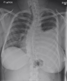
On exam, she has a very large goiter, and dullness to percussion over the left chest.


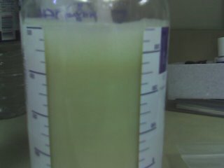
Her pleural fluid is shown here:

Thoughts?
5 comments - CLICK HERE to read & add your own!:
The fluid looks chylous. Maybe the tamoxifen was for a breast Ca and all that soft-tissue density in the CT are mets to nodes with lymphatic compression and a chylothorax.
Sarcoidosis and even the goiter itself can also cause chylothoraces.
I guess the first step of course is to get the chemical studies and cell analysis on the fluid.
The WBC's in the fluid were 2800 with 95% lymphocytes.
pH 7.79, LDH 126 (serum 155), protein 5.5 (serum 8.6), glucose 148.
Trigylycerides pending.
The pic of the fluid is pretty cool. I guess an empyema could look like that on a posted pic but the numbers don't support that. I will take my chances with a chylothorax for now.
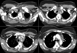
I agree-it looks chylous. The CT shows enlargement of some pre-vascular/peri-aortic nodes, and there's increased soft tissue in the superior mediastinum. Sarcoid is possible, but I'd be more concerned about a malignancy. I think a mediastinoscopy or even a Chamberland procedure is necessary to get tissue.
Post a Commenttest post a comment