77 year old admitted a year ago with pneumonia and referred for lung nodules. He had a PPD placed for unknown reasons in December, (no TB risks/exposures) that is 18 mm induration.
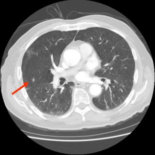
A CT is here: 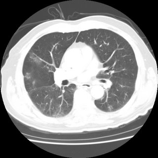 7 mm nodule
:
He has no respiratory or constitutional symptoms. Is there anyone that would
not do a BAL to rule out active TB prior to treating for latent?


5 comments - CLICK HERE to read & add your own!:
Those are very subtle findings... any mediastinal/hilar adenopathy? Any calcified granulomata? Are those little infiltrates present in other cuts?
There is a 7 mm nodule in upper lobe that is new. There is no adenopathy.
I added the nodule slice to the post.
Nothing calcified.
The rest of the CT is pretty unremarkable.
Does he have any old PPDs?
I don't think this is active TB. The radiographic changes are very subtle and if he had air-space dz with TB with no old calcified changes (hence, fairly acute disease) I would expect more symptoms. It sounds from your description that he doesn't even have a cough...
Now, having said that I think you are kind of stuck digging a little deeper: if you start INH for LTBI and those changes turn out to be atypical TB he will get a resistant bug. If you start 4 drugs for therapy and he is not coughing, you will not have any cultures and he will be stuck on 4 drugs with side effects for 6 months.
I would consider one of 2 things: a bronch as you suggested or, give him a quick course of "regular" ABTx and repeat a CT in 4 weeks. If it resolves or improves and he does not have symptoms of active TB you would have an alternative explanation and could go ahead with Tx for LTBI.
I like the bronch optin best because you can r/o active TB right away instead of waiting 4 weeks. I agree tht the pre-test prob for active TB is low, but with no known previous PPD (and now 18 mm) and a new 7 mm RUL nodule with some ground lass-ish in the middle, I thought it would be best to do a quick BAL...
I think a combination approach is reasonable. Do a bronch to get the samples, and re-image in 6 weeks if the bronch is negative. Then, treat for LTBI and, if the radiographic lesions progress, consider further diagnostic evaluation for non-TB disease.
Post a Commenttest post a comment