Showing posts with label lung lesions. Show all posts

Thursday, July 26, 2007

Steroid responsive adenopathy?

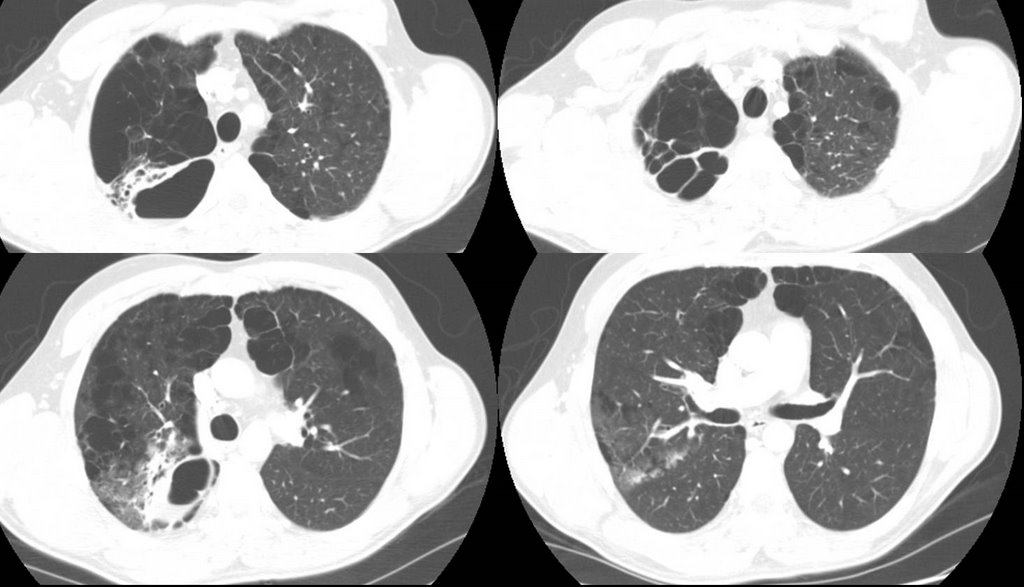
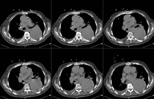
65-year-old woman presented initially for evaluation of shortness of breath. A CT thorax revealed some patchy airspace opacification RUL (see below) . She underwent bronchoscopic evaluation and it demonstrated some interstitial inflammation with hyperplastic type 2 pneumoctyes. No granulomas seen. No fungal or AFB elements on BAL. A cell count was not sent.
She was begun on a course of steroids 40mg and felt her breathing improved significantly while on them, but once she stopped them, her shortness of breath recurred. Her cough has also recurred, though it's mostly non-productive. A CT was repeated 5 monts later and is shown below.


Question: What ILD had adenopathy and is steroid responsive? The only 2 I can think of are sarcoid and berrylliosis, but the interstial inflammation would be inconsistent with that. Any thoughts out there?

Friday, April 13, 2007

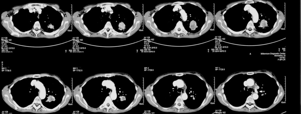
Cavitary mass

86 year old man with history of dementia brought by his caretaker for mental status changes and lethargy. He lives in an assisted living facility, but has no TB risk factors otherwise (He has his own apartment-like structure, but I beleive there is a common area).

His CT:


There was no hilar or mediastinal adenopathy.
The thickest wall area on that lesion was 4-5 mm.

Someone placed a PPD and it was 11 mm. It was reportedly negative 6 months ago.
He is not productive of sputum, so 3 gastric lavages were done and they were all negative. What would you do next? Are you worried about active tb? Here are my thoughts
1. How sensitive is gastric lavage for afb?
2. location of the cavitary mass is not where tb should be.
3. is he a ppd converter, or was he anergic or are we seeing a booster effect
4. His family does not want an aggressive w/u for cancer, but on the other hand one would not want to send someone home to hospice with active tb....

Wednesday, April 04, 2007

Lung mass(es)

Unfortunately, our computer is not letting me retrieve the PET images on this patient so I'll try and describe it without puppets.
This is a 71 y/o man with long TOB Hx but still good mechanics (FEV1~78%) with a new lung mass. He was found to have a 2.5-cm mass in the RML and had a CT-guided Bx which was + for adenoCa. He then had a PET and came to see us: the mass was obviously hot but he had FDG-avid subcarinal and L hilar nodes and a 1-cm FDG-avid LUL peripheral lesion. I bronch'ed him and sampled his nodes and got lots of giant cells and lymphocytes but no malignancy (from both sites). A LUL BAL was non-Dx.
Would you Bx the LUL separately? And if so how would you approach this (synchronous vs metastatic lesions, etc.)?

Friday, February 23, 2007

Digging for gold


This nice woman with mild asthma lost a tooth crown and aspirated it as she was about to have lunch.

Check out the CxR. It was lodged at the first branching of the R bronchus intermedius with the smooth side (the shiny top of the crown) up towards us. Unfortunately I was using an optic (as opposed to digital, I know they are all "optic") scope and couldn't take a picture. I did snap a picture of the crown after removal with the basket on the side.

We did not have a bronch basket handy so an OR gallstone basket was just the perfect sixe.

Thursday, November 02, 2006

Sticks and (bronchial) stones...


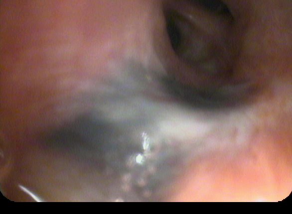
My partner bronch'ed this pleasant 78 y/o woman with no TOB Hx who had developed a persistent RML collapse. The first picture was taken while instilling NSS to keep the airway open.
The lesion was hard to biopsy and there was purulent mucus behind it.
The second set of pictures was a re-bronch 2-3 days later after he tried to remove as much as possible with the forceps.

Any suggestion, comments?

Tuesday, October 17, 2006

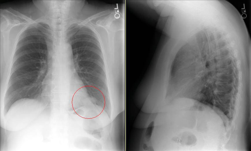
Abnormal CxR

I usually don't like those boards-type question that ask "what would you do next?" and only let you choose one option since we often do more than one thing in real life.
Having said that, I am curious as to what you would do next in this case. This is kind of a VIP patient so she was referred to a couple different specialists at the same time.
She is in her early 70s, quit smoking in 1972, near normal PFTs (FEV1~70%, normal DLCO) and has an abnormal CxR. It started as an URI and then she developed a "deeper" cough with purulent sputum production and some pleuritic CP.
The abnormal CxR (and some CT cuts) are shown below.

She had no adenopathy on the CT.
What would be your first step?

Wednesday, October 04, 2006

Dyspnea and a LUL mass

Here's a case from Arenberg:

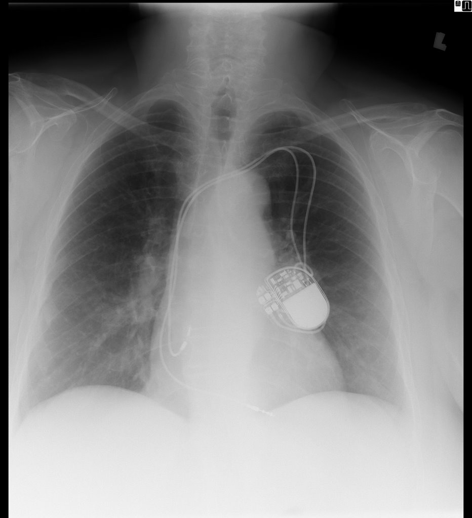
This is 69-year-old lady with coronary artery disease, diastolic congestive heart failure, and dyspnea on exertion. She came to see me because her cardiologist said she has been experiencing increased dyspnea out of proportion to her normal symptoms for ~3 months. Other than a pacemaker over her left chest, her plain chest x-rays were normal

and her pulmonary function tests were mildly restrictive (FVC ~69%), actually improved from her last testing ~ 4 years prior.

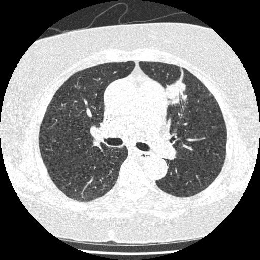
I got a VQ scan that was normal, but only on her second visit (when I actually asked) did I learn that she has had 2 birds as house pets for the last 3 years. She further revealed that she has felt unwell going back about 2-1/2 years. One of the birds was in fact sick, and during this time, she fed the bird from mouth to mouth. Yes, I am serious. She was DEAD SET against getting rid of the birds, so I got a CT scan in anticipation of doing a bronch, to prove she had HP.

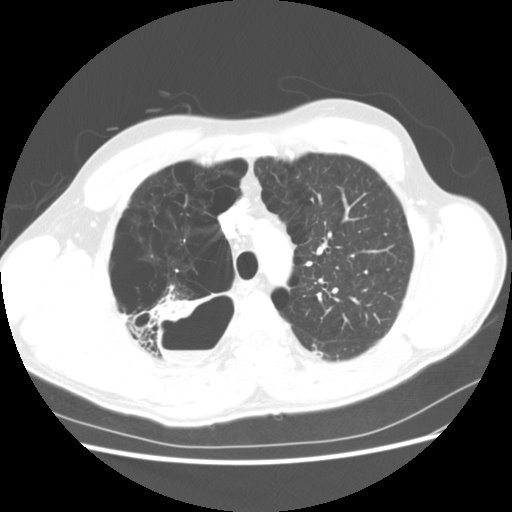
The CT showed very subtle changes of ground glass, and was actually read as no evidence of ILD, but surprise surprise, she now had a LUL mass that was not seen on the CXR taken 2 months earlier. The radiologist thought it was likely to be inflammatory. What would you do?

I have more images, and some cool pictures to follow, but I’ll hold onto them until people have a chance to comment.


--------------------------
UPDATE:
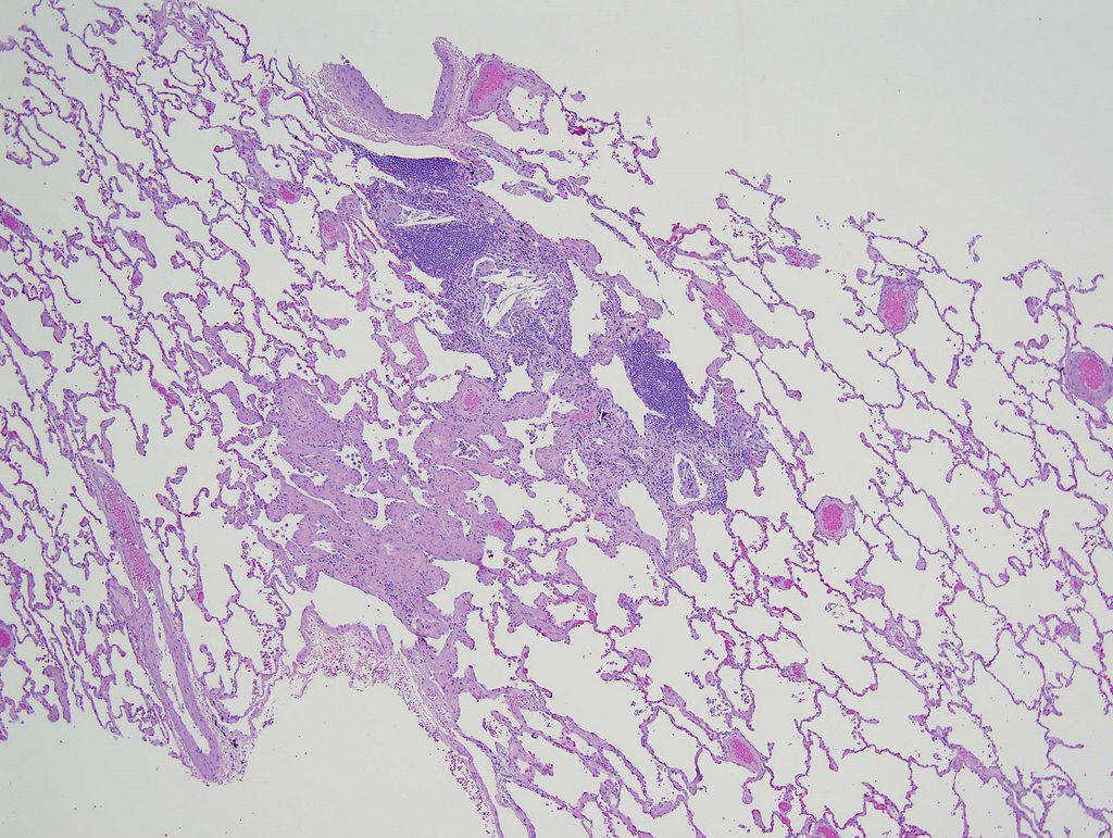
"...good evidence for chronic HP in the form of a patchy chronic
bronchiolitis that includes occasional multinucleated giant cells of the
sort commonly seen in that condition"

Comments from our pathologist.

Tuesday, September 26, 2006

fluid filled lesion

"Anonymous" sent us the following CT slices of a 38 year old who presented with cough and fever. AFB were negative x 3. He was not very productive of sputum and what they did get had many epithelial cells.
How would you proceed if this was your patient?

Update on Mass and Infiltrates

I had posted on this 59 y/o woman with a funny presentation: she had had an episode of syncope at home, hit her head and had a skin lac requiring 7 stitches. While being worked up in the ER they found her to be hypotensive and febrile with an abnormal CxR and CT of the chest. She had MSSA in her sputum and a dense lung "mass". I bronch'ed her and got "organizing pneumonia" but no malignancy. She got ABTx and the "mass" went away completely. She did however had persistent smallish alveolar infiltrates seen below:

I re-bronched her focusing on the infiltrates since they had persisted for 3 months after the initial presentation. She had a lot of eosinophilic proteinaceous alveolar material and our pathologists sent the tissue to T. Colby who thought this was PAP.
Would you be satisfied with this Dx on TBBx or would you pursue an OLBx?
How about testing for anti-GM-CSF Antibodies?
She has almost normal lung function, how would you treat her?

Monday, September 18, 2006

Follow-up to Pulmonary-thyroid

This is the patient with the hot pulmonary nodule and lung mass.
We decided to start with the thyroid and a bronch. The thyroid mass (it was not cystic on CT) was a follicular carcinoma with local invasion. No lymphnodes and the other lobe was clean.
The bronch was non-diagnostic and radiology felt the mass was too low and there was too much lung in its way for a good CT-guided needle.
She went to see our CT surgeons and we figured out why the bronch was non-diagnostic: it was not a lung mass but a diaphragmatic mass. It was an aneuploid adenomatoid tumor still not clearly characterized. Incidentally, there was a second focus of this same adenomatoid tumor in the superior segment of that LLL, which had not been seen on CT or PET (at ~7mm).
What do you think?

Wednesday, September 13, 2006

The pulmonary-thyroid connection


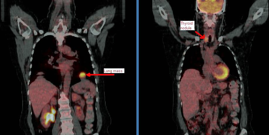
This patient came to me pre-worked up so I will show all the data she had had thus far. This is a 65 y/o woman with no TOB Hx, normal PFTs who had a persistent cough. She had an abnormal CxR (see above) and here PCP ordered a CT and then a PET-CT and she was sent to us.
Her PET scan showed two very FDG-avid lesions. A L basilar lung lesion and a R thyroid nodule with no other abnormal uptake. She is euthyroid clinically and by TFTs.
Do you think the lesion are related?
Would you biopsy one, the other, both?

Tuesday, September 12, 2006

LUL lesion, still undiagnosed

70 year old woman with COPD FEV1 34% predicted, but she is well compensated and on no supplemental oxygen (91% on RA). A LUL lesion was noted (see below) and workup included 2 bronchs (one with ultrasound-guided) and 1 CT-guided biopsy. All were negative for malignant cells. BAL was not done but Tbbx showed Atypical lymphoid cell with acute inflammation and Filamentous bacteria. BAL was not sent so we don't have micro ID (the bacteria was identified by path).


Cancer is obviously still number one on the list, but I dont think she's a surgical candidate and in terms of treating with chemo, we still dont have a tissue diagnosis. On the other hand, the filamentous bacteria finding is interesting. What is your impression and what would you all do next? Thanks.

Monday, August 28, 2006

35 year old with cough, chills

35 year old without PMhx except a MVA 5 years ago, anxiety and depression. Now with chills and subjective fever and weight loss for the past few months. Meds: Paxil. Fhx: brother died of liver failure, patient does not know etiology. SH denies IVDA. 2 PPD x 20 years. Quit 5 yrs ago. HIV negative. Exam Afebrile. VSS. Some decr breath sounds upper lung zones b/l. WBC 11.


What would you do next?

Tuesday, August 08, 2006

Mass and infiltrates

This is a 59 y/o woman with a funny presentation: she had an episode of syncope at home, hit her head and had a skin lac requiring 7 stitches. While being worked up in the ER they found her to be hypotensive and febrile with an abnormal CxR and CT of the chest. She had MSSA in her sputum and the dense "mass" seen on the left images. I bronch'ed her and got "organizing pneumonia" but no malignancy. She got ABTx and the "mass" went away completely.
She has felt better and has good PFTs. However, it has now been 3 months and the peripheral faint patchy areas of reticular disease remain there and unchanged.
She has only minimal DOE. She quit smoking in 1988 and has no unusual exposures.
How would you pursue this little infiltrates? (they are present in the lower lobes as well).

Wednesday, June 07, 2006

left lower lobe lesion. What would you do next?

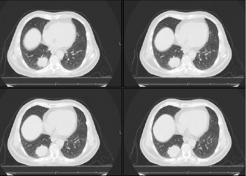
67-year-old Caucasian female who was a previous RN, presents because prior to getting her vascular surgery, she was having a double aortofemoral bypass for claudication. Had a chest x-ray done showed an abnormal opacity on the left lower lobe. She says that she has had worsening shortness of breath for one and half years and has
really had any cough. Denies any fever, chills, rigors, chest pain, or weight loss. Denies any hemoptysis. Denies any previous infections. Denies any headache, nausea, vomiting, or diarrhea.

AST MEDICAL HISTORY: Peripheral vascular disease, hypertension, hyperlipidemia, previous history of smoking.

MEDICATIONS: Atenolol, Protonix, Procardia, hydrochlorothiazide, Lipitor, TriCor, aspirin, multivitamins, and Tylenol 3.
SOCIAL HISTORY: 50-pack-year smoking history, stopped 2 years ago. No ETOH or drug.
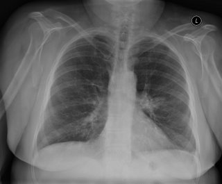
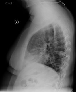
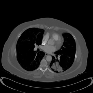


No mediastinal or hilar adenopathy.
Repeat CT 1 month later showed no change in size.

Patient was bronched with non-diagnostic tissue.
Cytology brush from left lower lobe was negative for malignant cells.
Micro on BAL: 30000 CFU per ml alpha-hemolytic Streptococcus, 8000 CFU Neisseria; 9000 CFU Micrococcus; 5000 CFU Streptococcus, non-hemolytic.

What would be your next step (or any other questions you have)?

Wednesday, April 26, 2006

Follow up to Nodule and Mass

I had posted this but didn't get much feedback. This is the fairly healthy 40 y/o woman with some chronic back pain but no pulmonary or cardiac disease referred to us for a "new" pulmonary mass. She has never smoked and has had no pulmonary symptoms. She had been told about 10-15 years ago that she had a calcified R lung nodule and that has just been followed along. However she had had recent imaging for her back and they found that mass in the azygoesophageal recess.
Jennings astutely pointed out that the posterior mass looked very heterogeneous and almost cystic. The films were outside films so we decided to try a bronch and a repeat CT. Our radiologists felt comfortable that the peripheral lesion looks like a hamartoma and agreed the mass appeared cystic like a bronchogenic cyst. The bronch (we tried some FNA passes. Was fairly unremarkable and essentially non-diagnostic.
Would you have done something else? Would you consider a PET? If negative we might not repeat imaging studies as frequently... Or would you even bother with scheduled CTs if she remains asymptomatic?

Saturday, April 15, 2006

Follow-up to What is your algorithm?


This is the 64 y/o man with significant TOB Hx and a lung mass.
I had the same thoughts as Jeff H.
His FEV1 incresed to >2L (>60%) on Spiriva. A bronch revealed no endobronchial lesions but was otherwise non-diagnostic. The PET lit up up (brightly, see above) only on the mass and nowhere else.
What would you do next?

Monday, April 10, 2006

Abnormal CT scan


We were asked to see this delightful 67 y/o woman with no respiratory symptoms for an abnormal CT scan of her chest. She has had some back pain and a nodule was found on a CxR. The CT scan above was obtained and she was referred to us. She quit smoking in 1992.
She has good lung function with normal FVC and FEV1~ 70%.
She was set up for a PET scan and the images are seen below:

I don't know if the writing projects well but the RUL lesion is positive and she has positive paratracheal and R hilar nodes. There was also a positive 4-mm R supraclavicular node that did not project well.
What would you do next?

Wednesday, April 05, 2006

Unexpected findings

A 59 year old man was referred for an unexpected finding in the RUL discovered during an operative airway exam prior to resection of a large left lung mass. The lung mass turned out to be a solitary fibrous tumor. We were asked to evaluate the unexpected lesion in the right upper lobe. At bronchoscopy, I saw a lesion at the very proximal anterior segment of the RUL.


This was adherent and non-mobile. The patient is otherwise healthy, and his immune-system is intact.

Thoughts?

Wednesday, February 15, 2006

Follow-up to Wednesday Radiology - Case 2

'Can't hide anything from these smart people.
Check out the CT (unenhanced because Creat is 2.0). We did not feel like biopsying the "mass" either...