Showing posts with label mediastinum. Show all posts

Thursday, July 26, 2007

Steroid responsive adenopathy?

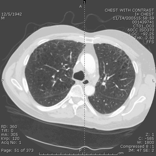
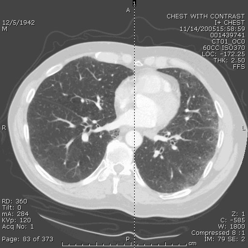
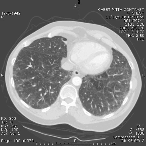
65-year-old woman presented initially for evaluation of shortness of breath. A CT thorax revealed some patchy airspace opacification RUL (see below) . She underwent bronchoscopic evaluation and it demonstrated some interstitial inflammation with hyperplastic type 2 pneumoctyes. No granulomas seen. No fungal or AFB elements on BAL. A cell count was not sent.
She was begun on a course of steroids 40mg and felt her breathing improved significantly while on them, but once she stopped them, her shortness of breath recurred. Her cough has also recurred, though it's mostly non-productive. A CT was repeated 5 monts later and is shown below.


Question: What ILD had adenopathy and is steroid responsive? The only 2 I can think of are sarcoid and berrylliosis, but the interstial inflammation would be inconsistent with that. Any thoughts out there?

Monday, March 19, 2007

Mediastinal adenopathy, hypoxia

Dr.Carrillo submitted a case for your enjoyment/pontification:

49yo Hx of ETOH and presumptive cirrhosis. Exposed to birds through his early years. 2000 had abnormal CT thorax with interlobular septal thickening, Pleural thickening encasing the lungs and multiple adenopathy in the med. Mediatinoscopy report says multiple venous collaterals may be early SVC.Thickened fat through the mediastinum.Bx of LN neg,no Cx sent. 2003 - Same CT findings.

Now presents with hypoxia and CT findings similar as before. The radiologist describes that the mediatinum looks abnormal with strading. I think this guy probably has histo with some fibrosis of the medistinum. I wonder if he has compression of the thoracic duct with causing his lymphatics to be prominent and show like interlobular septal thickening on CT and he had previous Chylo?. I was going to do broncoscopy with TBBX and TBNA and serologies. Any other idea or opinion about this case. Do you think I should further investigate the possible thoracic duct obstruction?

Thursday, July 27, 2006

mediastinal nodes

Case submitted from outside:

63 year old smoker with hypoxia. CXR looks extremely hyperinflated. PFTs with ratio of 0.67, FEV1 87% predicted and FVC 93%. Lung volumes normal except for an inspiratory capacity of 73% predicted (volume done by helium dilution). DLCO 35% predicted. CT scans included below and are basically unchanged from 7 months ago. Mild mediastinal LAD, with the largest being approx 2cm. Also stable over 7 months. Would anyone pursue a biopsy of the nodes or the lung? Other thoughts?


Tuesday, June 13, 2006

Follow-up to Radiology Thursday

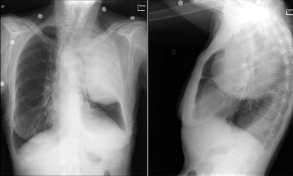
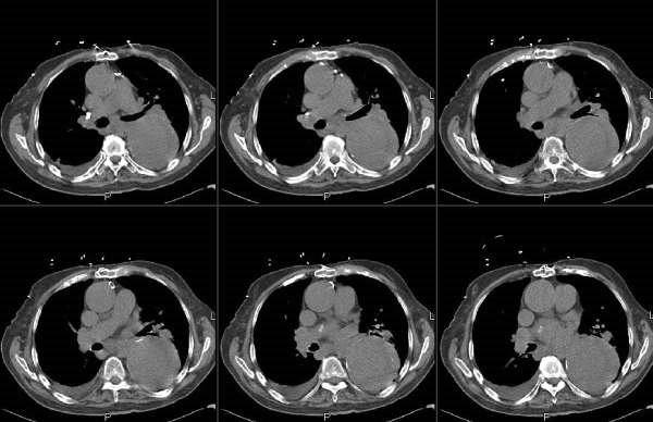
Jennings picked up on the Left mediastinal mass. The patient could not stand up for the lateral so a CT was obtained:

Monday, June 12, 2006

Mediastinal adenopathy

This case was submitted by one of our readers:
"My husband had a CT-scan and an enlarged mediastinal node (1.5 cm.) was noted in the short axis adjacent to the descending thoracic aorta. At the time, he had suffered an episode of pain and shortness of breath, and was diagnosed with pleurisy. One month later, a repeat CT scan showed that the mediastinal node was still enlarged, though had not increased in size. What is the normal protocal for monitoring this condition?"

Friday, May 12, 2006

Lung cancer and adenopathy

A reader submitted this question:
A patient was just diagnosed with lung cancer and will be undergoing surgery soon. The tumor was discovered during an incidental Cxray and is approx 2.5 cm and located in the right lower lobe. We were encouraged to find that the ct scan showed no spread to the the lymph nodes and no pleural effusion. However, a subsequent PET scan showed bilateral Hilar uptake but no mediastinal uptake. The Thoracic surgeon scheduled a medianoscopy before the lobectomy just to make sure there was no spread of the cancer to the lymph nodes. His feeling is that the PET scan would have shown some mediastinal uptake on the PET scan and feels that the bilateral Hilar uptake is probably not cancer.
Just wanted to get some other views on this situation.

Monday, April 17, 2006

Symmetric bilateral hilar adenopathy

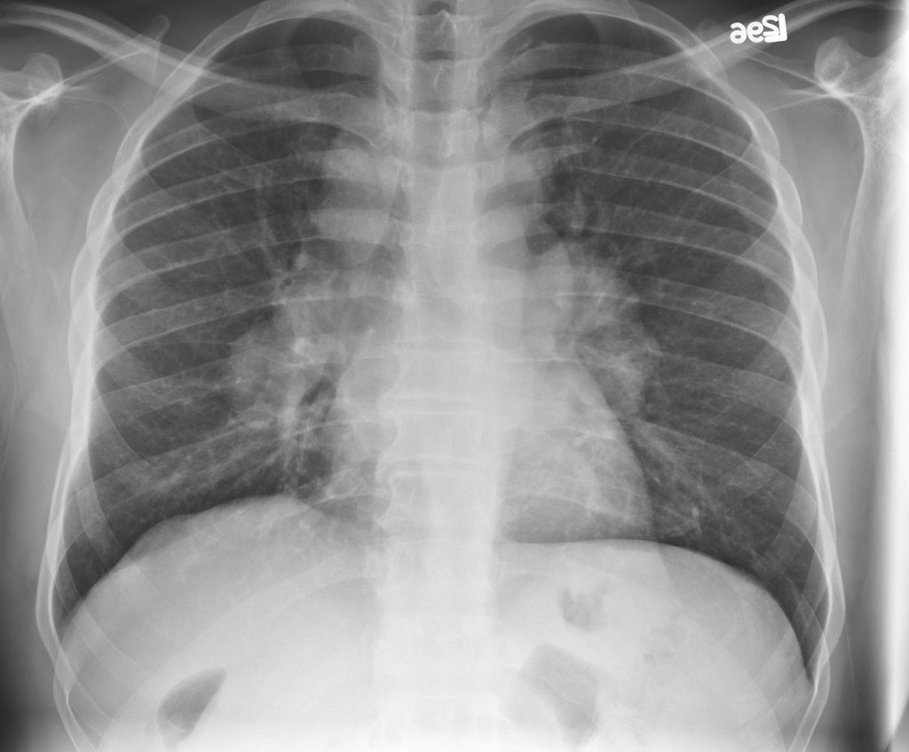
47 year old African-American man who was asymptomatic at the time of this chest xray:


His PFT's were normal. LFT's were slightly elevated (Alk phos 192, GGT 152) with a normal ultrasound and no abdominal complaints. A presumptive diagnosis of sarcoidosis was made and the patient was simply followed. 5 months later he c/o subjective fever and night sweats. No respiratory symptoms. A chest xray was actually improved:


What would you do? Are you still satisfied with the diagnosis of sarcoid? (I am aware of a study that found that in almost all cases with symmetric adenopathy, the dx is sarcoid.) WOuld you want to definitely dx it or rule out other causes?
If you are satisfied with the dx, how would you handle the constitutional symptoms? Assuming f/u PFT's are not in decline, would you treat these B symptoms with immunosuppressive therapy?

Wednesday, March 08, 2006

Abnormal CxR


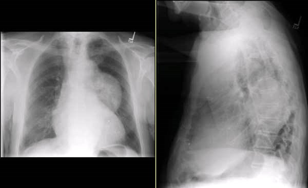
This is the 79 y/o mother of one of our physicians. She has a remote TOB Hx (quit >15 yrs ago) and subacute dyspnea and fatigue (a couple of months'). She presented through the ED with dyspnea and L atypical chest pain. Her nasal swab was positive for influenza and she had the CxRay seen above. What is your DDx and what would you do next?

Wednesday, February 15, 2006

Follow-up to Wednesday Radiology - Case 2

'Can't hide anything from these smart people.
Check out the CT (unenhanced because Creat is 2.0). We did not feel like biopsying the "mass" either...

Radiology Wednesday - Case 2


We were called by one of the cardiologist for this abnormal CxR. How would you approach the radiographic abnormality?