Showing posts with label Pulmonary nodules. Show all posts

Tuesday, February 27, 2007

SPN

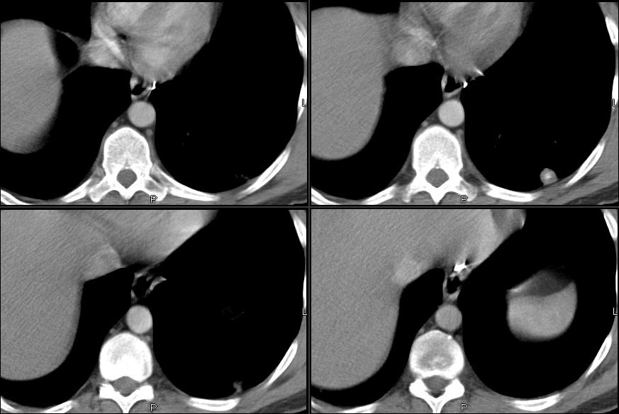
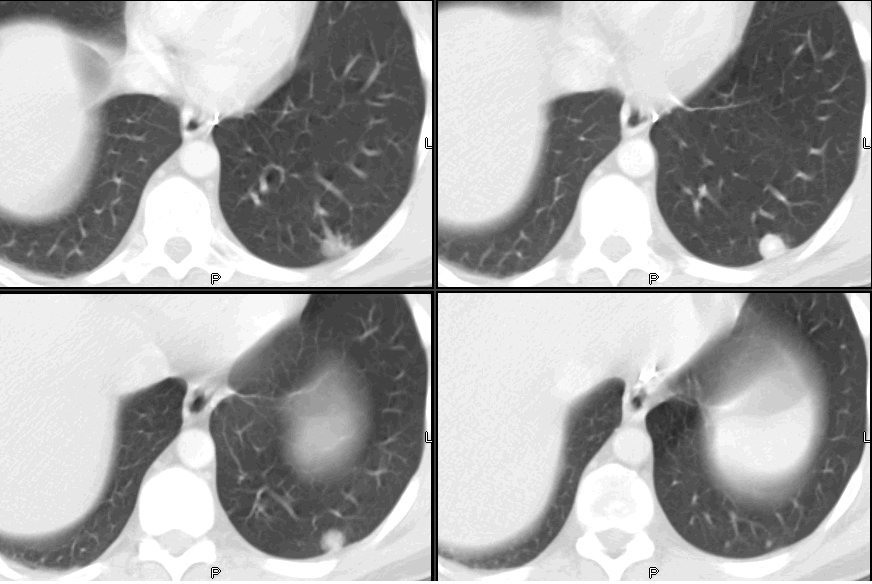
79 year old female non smoker presented with hypercalcemia and a low PTH. She was pan-ct'd and all that came up was this SPN in the RML:

It's size is about 7 mm. Of note, the CT slices were 7 mm.
How would you proceed?

Thursday, January 18, 2007

Solitary pulmonary nodule

I recently saw a 62-year-old woman with a past medical history of fibromyalgia who received a CT scan for chest pain, which picked up an incidental pulmonary nodule.
She is asymptomatic; no shortness of breath or cough. She is a 35 pack-year smoker and has not been able to quit. Her spirometry is normal.
A CT is shown here


What would you do?
1. refer to CTS for resection
2. Get a PET (and what would you do with that info?)
3. Follow radiographically, say, with a repeat CT in 3 months?

I'll tell you what I did after comments.
Feel free to enter your name as "anonymous".....

Monday, January 15, 2007

multiple pulmonary nodules in system lupus

Case from slkanowitz:

57 y.o. white female with SLE for 20+yrs presents with multiple small non calcified nodules on chest CT. CT was done for evaluation of asthma. Pt. symptomatic only with her usual migratory pleuritic pain associated with her lupus, and mild, intermittent asthmatic symptoms present since young adulthood. Asthma is treated prn with albuterol inhaler. Largest nodule is 6mm, most are peripheral and they are bilateral. Follow up chest CT scan 4 mos. later shows resolution of some of the nodules, the remaining ones show no growth. PFT are normal, O2 sat. normal. There has been no change in symptoms and no change in treatment for her lupus during that time. Other pertinent medical problems include allergic rhinitis, chronic sinusitis, S/P antral windows, and chronic bronchitis. Negative for pneumonia, SLE pneumonitis, RA, ANCA, anti-phospholipid AB. Recent echo WNL. No hx malignancy.
Hx significant for living in Ohio River Valley prior to 1973 and Southern Calif. during 1990, but no history unusual respiratory infections.

1998 chest xray normal. No xrays done until CT scan of chest and sinuses in 09/06.

SLE confirmed without evidence of other connective tissue disease. Pt. on Placquenil 200mg BID and Methotrexate 22.5 mg/week.


She did not pose a question but I assume it is what to do about the multiple nodules.
Since the nodules decreased in size over 4 months, this is likely inflammatory and a repeat CT in 6 months for that last nodule would be what I would do. I'm not sure what her smoking history is, but pretest prob for malignancy is low here. Any one else wish to comment?

Saturday, December 09, 2006

Follow-up to Pulmonary Nodules

This is the 51 y/o woman with COPD and pulmonary nodules.
We had the same concerns and DDx as proposed in the discussion. I did a bronchoscopy and we got good samples consistent with RB-ILD.
She was counselled again regarding TOB cessation and agreed to try varenicline. She will follow-up in a few weeks and we will monitor her CxR and PFTs.

Tuesday, December 05, 2006

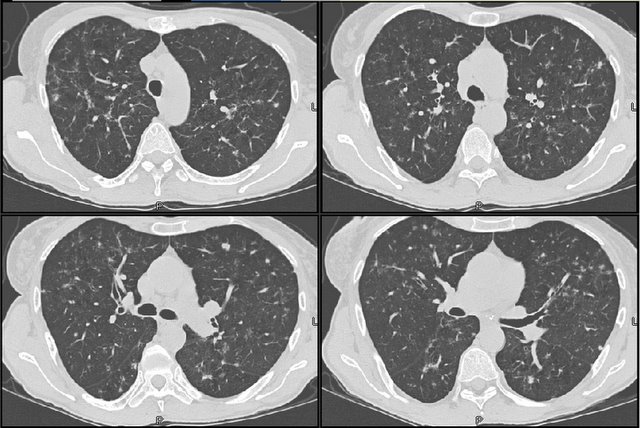
Pulmonary nodules

This is a 51 y/o woman followed in our office for moderate COPD. She continues to smoke and has had worsening dyspnea and weight loss for a couple months. Her CxR and CT scan, which in the past showed hyperinflation and nothing else, now have these pulmonary nodules.
What is your DDx and wht would you do next? Posted by Picasa

Tuesday, November 14, 2006

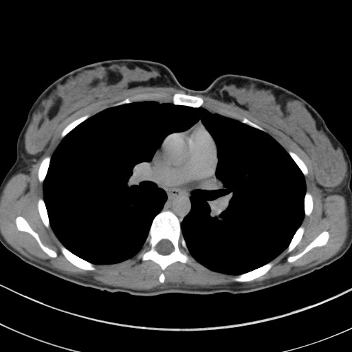
LUL nodule with ipsilateral LAD

25 yo female with no PMH presents with above.
She was well until April when she travelled to Phoenix to visit friends. When she returned, she had a bad URI. Her PCP obtained a CXR which showed a shadow in the L hilum, and a CT was recommended.
The CT was from an OSH and resembles the CT shown below. The L hilar fullness is a node clearly visualized in the OSH CT; the CT I show is without contrast (she got hives after the first CT). The rec on the CT was to obtain a repeat in 3 months. This is the 3 month CT.

She is currently asymptomatic other than a 20 lb weight loss since 4/06. She has not changed her eating habits or exercising habits.

PMH: Non-smoker but moderate second-hand smoke from her parents.


Labs: CBC, chemistries, U/a are all normal.

What would you do?

Monday, September 18, 2006

Follow-up to Pulmonary-thyroid

This is the patient with the hot pulmonary nodule and lung mass.
We decided to start with the thyroid and a bronch. The thyroid mass (it was not cystic on CT) was a follicular carcinoma with local invasion. No lymphnodes and the other lobe was clean.
The bronch was non-diagnostic and radiology felt the mass was too low and there was too much lung in its way for a good CT-guided needle.
She went to see our CT surgeons and we figured out why the bronch was non-diagnostic: it was not a lung mass but a diaphragmatic mass. It was an aneuploid adenomatoid tumor still not clearly characterized. Incidentally, there was a second focus of this same adenomatoid tumor in the superior segment of that LLL, which had not been seen on CT or PET (at ~7mm).
What do you think?

Wednesday, September 13, 2006

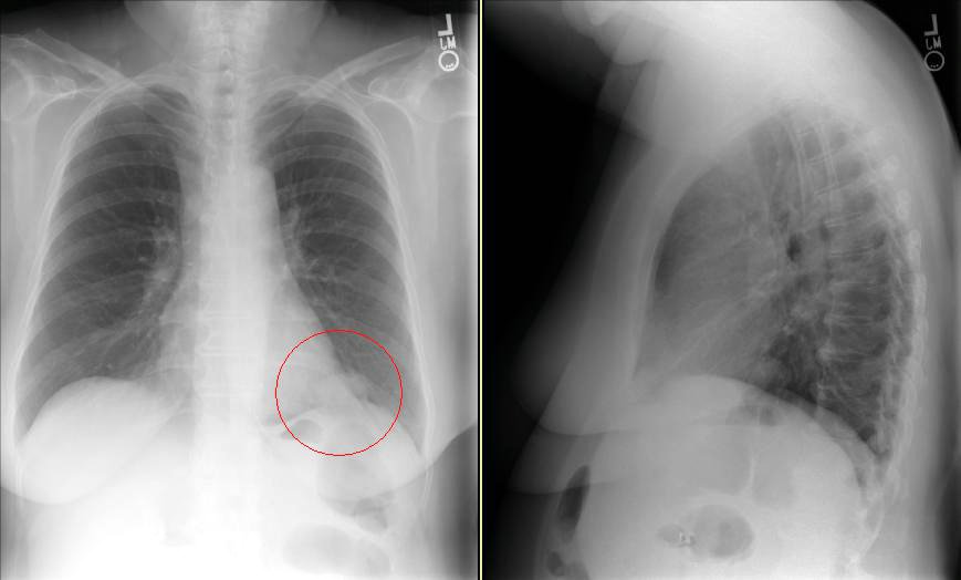
The pulmonary-thyroid connection


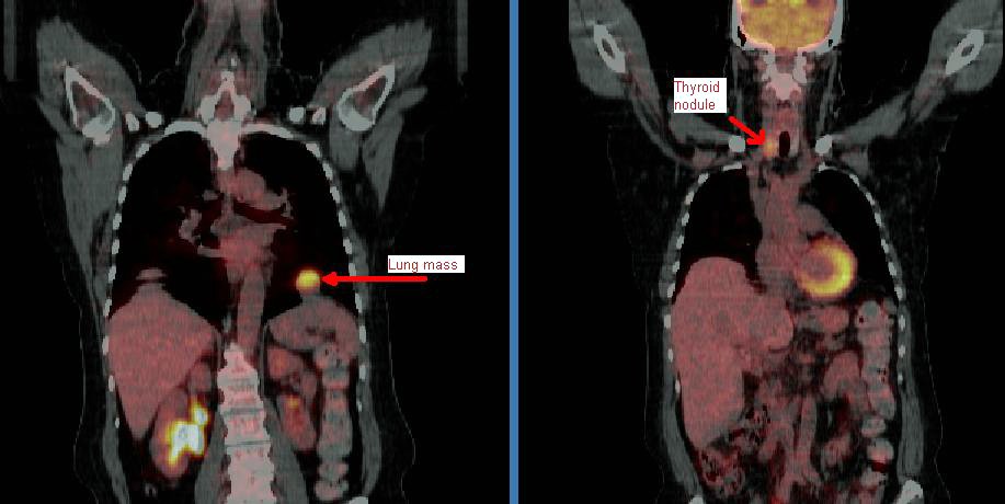
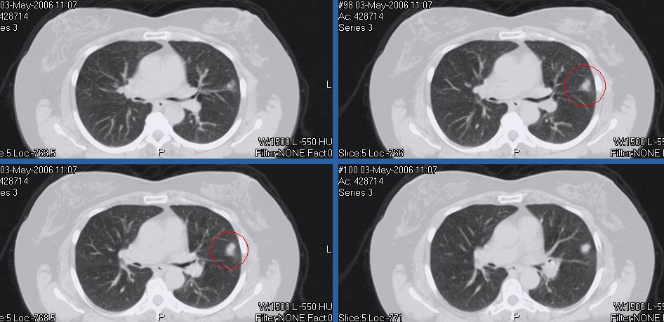
This patient came to me pre-worked up so I will show all the data she had had thus far. This is a 65 y/o woman with no TOB Hx, normal PFTs who had a persistent cough. She had an abnormal CxR (see above) and here PCP ordered a CT and then a PET-CT and she was sent to us.
Her PET scan showed two very FDG-avid lesions. A L basilar lung lesion and a R thyroid nodule with no other abnormal uptake. She is euthyroid clinically and by TFTs.
Do you think the lesion are related?
Would you biopsy one, the other, both?

Wednesday, September 06, 2006

Multiple pulmonary nodules

The patient is a 69-year-old African-American woman who came to pulmonary clinic for multiple pulmonary nodules that were picked up in the course of abdominal CT for workup for abdominal pain with increased LFTs (which have since normalized). She has a history of hypertension, diabetes, peripheral vascular disease, and hyperlipidemia. She is a nonsmoker. No fever chills cough SOB, diaphoresis or constitutional symptoms.
Exam: A pleasant woman in no acute distress. WT of 197, BP 185/83, P is 65, R 18, O2 sat is 97% on room air. Trachea midline. No lymphadenopathy and no supraclavicular adenopathy. No thyromegaly. Lungs: Clear to auscultation. No wheezing, rales, or rhonchi. No dullness to percussion. No egophony, symmetric chest wall on inspiration. Heart: Regular rate. S1, S2. Abdomen: Positive bowel sounds. No masses. No liver edge felt. No clubbing, cyanosis, or edema.

Data:
PFT's: FEV1/FVC 81, FEV1 76% predicted, FVC 73% predicted.
CBC with hemoglobin of 10.3, MCV of 94.6. Plts normal. Lytes-7 normal. ANA and rheumatoid factor negative
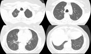


These nodules are all non-calcified. The largest is 8 mm. There is interval growth of left lower lobe nodule and right apical nodule compared to a CT 6 months prior. The subcarinal soft tissue is "prominent" but ambiguous whether it's enlarged.

On abd CT, the spleen, pancreas, kidney, gallbladder and adrenals are unremarkable.
2 calcified hepatic granulomas are seen.

Any thoughts on workup or management?


.

Monday, August 14, 2006

Outside case: Adenopathy

This was submitted to us:
34yr.old female w/ previous rt low lobe nodules since 2001 CT revealed the nodules while patient had chickenpox.
New CT in 2006 shows new node on lt. upper diaphragm. Last CT 6mo. prior didnt show nodule, which is uncalcified unlike rt. lung nodes.
PT. has nightsweats wt. loss chronic pelvic abdominal pain jointpain/swell/stiff no appetite ab.bloat chronic urinary symptoms and UTI.
Now with SWOLLENLYMPHNODES W/LYMPH SURFACING AND RUPTURE ON NECK BACK OF HEAD BEHIND EAR. PT. is 11/2 ppd smoker of 13yrs. PET done 4/06 and no malignant chest activity. labs almost normal.

Sunday, July 30, 2006

Pulmonary nodules

This is a 44 y/o man who works here in the hospital. He is a ctually a nice guy so I'm trying not to find anything too interesting on his case.
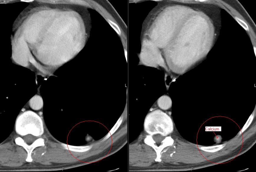
He is very healthy and smoked a pack-a-day for ten years but quit in 1988. He had travelled to the gul area and came back with a severe gastroenteritis. It seems that it was so bad the primary team got a CT of his abdomen. He did OK but the lower cuts of the lung revealed a ~1-cm partially calcified LLL nodule. He is completely asymptomatic from a respiratory perspective. His PFTs are completely normal. We got a dedicated CT of the chest and found the following nodules: (a 9-mm RUL a ~5-mm L apical and the same LLL).
The RUL has a little bit of excentric calcium as does the LLL one. Would you PET, biopsy, watch or else?

Tuesday, July 25, 2006

New lung nodule

This is a 72 y/o man with a significant previous TOB Hx, moderate COPD who was referred to us for a new pulmonary nodule. He has had multiple urologic surgeries including a nephrectomy for transitional cell ca (it seems he had a "high" transitional ca of the calicial system). He has a new nodule on the contra-lateral (remaining) kidney and had a new CT and a pulmonary nodule was found. This is a LUL non-calcified nodule with no air-space disease and no thoracic adenopathy.
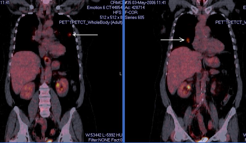
A bronch was non-diagnostic and a PET (see below) only showed intense uptake on the nodule and nowhere else (the remaining kidney had its usual physiologic uptake).
Do you think the two lesions are related? Would you try and FNA the lesion? His PFTs would tolerate a lobectomy. Would you just go straight to surgery?

Thursday, June 01, 2006

Follow-up to lung nodules


This is the woman with B/L pulmonary nodules. I was also concerned about these non-calcified nodules so she had a PET scan and we performed a bronch. Both nodules lit up on PET and she had some faint, low-SUV uptake in the mediastinal nodes. The TBBx of the R lesion were + for a non-small-cell lung Ca.
She has good PFTs.
What would you do next?

Tuesday, May 30, 2006

Lung nodules

We are back from ATS and will start posting new cases.
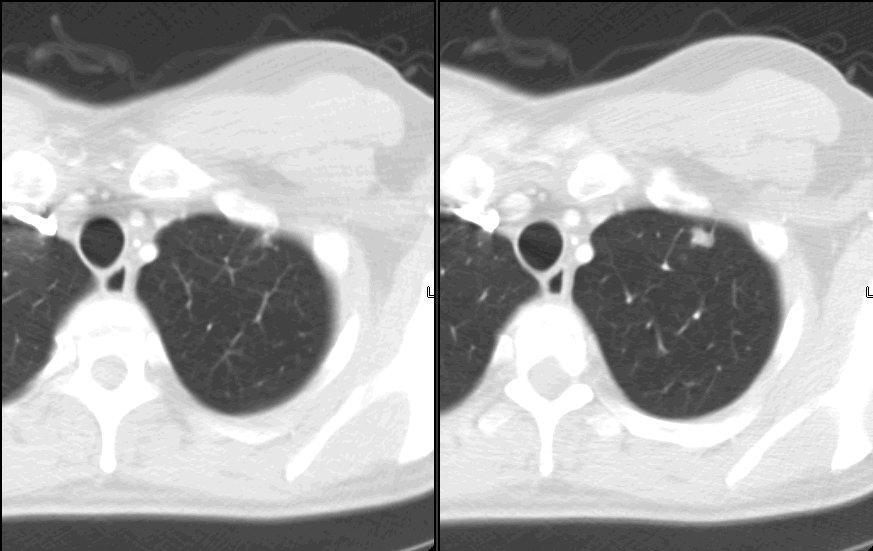
This is a 60 y/o woman with a significant previous TOB Hx with very little on the way of symptoms. She had some cough and a CxR revealed a small nodule on the R. The CT scan below actually revealed two nodules, one on each side (see below, they are both fairly small). How concerned would you be and how would you work her up?

Wednesday, April 26, 2006

Follow up to Nodule and Mass

I had posted this but didn't get much feedback. This is the fairly healthy 40 y/o woman with some chronic back pain but no pulmonary or cardiac disease referred to us for a "new" pulmonary mass. She has never smoked and has had no pulmonary symptoms. She had been told about 10-15 years ago that she had a calcified R lung nodule and that has just been followed along. However she had had recent imaging for her back and they found that mass in the azygoesophageal recess.
Jennings astutely pointed out that the posterior mass looked very heterogeneous and almost cystic. The films were outside films so we decided to try a bronch and a repeat CT. Our radiologists felt comfortable that the peripheral lesion looks like a hamartoma and agreed the mass appeared cystic like a bronchogenic cyst. The bronch (we tried some FNA passes. Was fairly unremarkable and essentially non-diagnostic.
Would you have done something else? Would you consider a PET? If negative we might not repeat imaging studies as frequently... Or would you even bother with scheduled CTs if she remains asymptomatic?

Monday, April 24, 2006

Pulmonary nodule

This 44 y/o man is one of our former nurses. He had gone to Florida on vacation and came back with an infectious diarrhea. The ER did a CT scan of the abdomen and they found a pulmonary nodule on the lower lung cuts (see below).
He has no respiratory symptoms whatsoever. He quit smoking in 1988 and had about a 10 pack-year total.
What do you think of the nodule (it is 11x12 mm) and what would you do next?

Tuesday, April 18, 2006

Nodule and mass


This images are from an outside radiology service so the quality suffered a bit when I copied it...
This is a fairly healthy 40 y/o woman with some chronic back pain but no pulmonary or cardiac disease referred to us for a "new" pulmonary mass. She has never smoked and has had no pulmonary symptoms.
She had been told about 10-15 years ago that she had a calcified R lung nodule (left pics) and that has just been followed along. However she has had recent imaging for her back and they found that mass in the azygoesophageal recess (right pics).
Would you be concerned? Do you think the two lesions are related and what is your differential?

Monday, March 06, 2006

Pulmonary nodules.

I had been following a 79 y/o man with small non-calcified nodules with serial CT scans. He is a lifelong non-smoker and one of his noudles was actually calcified and he had a calcified small hilar node. His nodules had been stable for about a year and with the lack of smoking Hx and other finding I was quite confident these findings were benign.
He developed a new URI and through his PCP, had a new CXR followed by a CT scan (see below). The CT revealed a small new left nodule. All the other previous findings had remained unchanged.

He then had a PET-CT: the nodule, and actually all the nodules as well, was PET-negative. However, the left hilum (not much there on CT was positive with an SUV of 2.54 and there was a positive subcarinal node with an average SUV of 2.2.
Are you very impressed by those numbers or would you write it off as inflammation and do a CT follow-up?

Tuesday, February 28, 2006

More pulmonary nodules


We see a lot of pulmonary nodules here in TN.
This is a man in his late 60s with severe COPD (FEV1~40%) who had some nodules on his CxR. He had a CT scan done and has 2 main nodules on the right (pictured) and a very small one (not pictured) on the left. His bronch was non-diagnostic and his PET was negative. Would you pursue this any further or just watch?