Showing posts with label airspace diseases. Show all posts

Wednesday, December 19, 2007

41 year old with HIV and fever

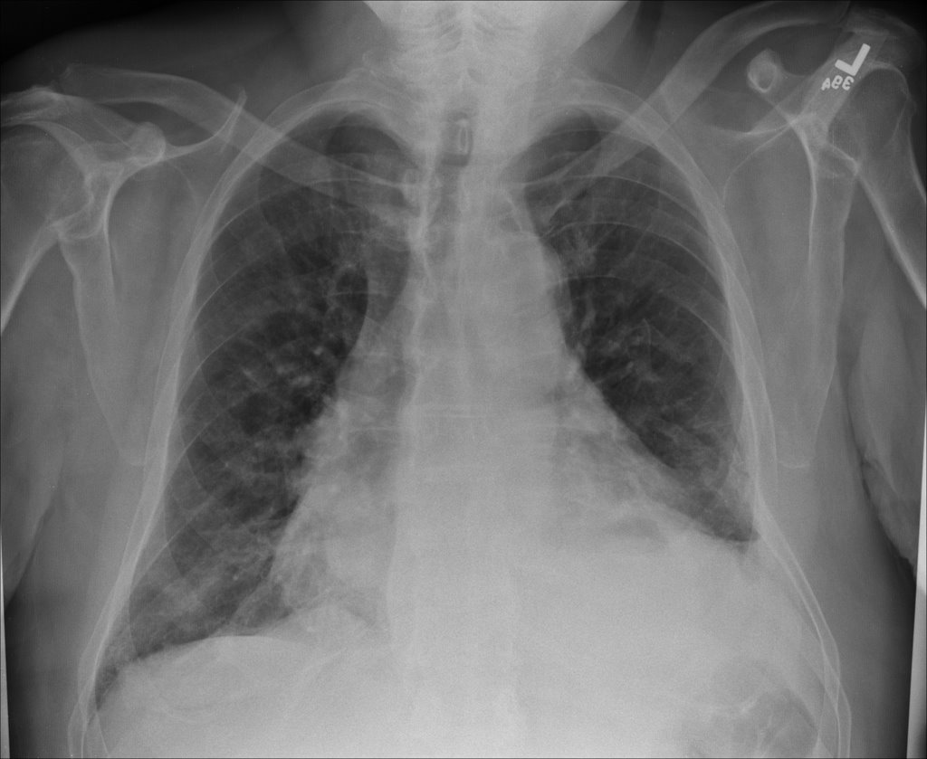
This is a 41 year old man with AIDS, CD4 below 10, who presented with shortness of breath and fever. He was admitted to a general ward but trasnferred to the unit a few days later for tachypnea. On the general floor, his vitals were normal except for some tachycardia. He was 97% on 2 l NC. CXR:


a full workup was done to look for infectious source.
sputums: AFB negative x 3, fungal negative. PCP negative (no PCR, no bacteria
CSF - negative for infection.

What kinds of things could be causing these findings and what would you do?

Wednesday, September 05, 2007

Pulmonary infiltrates


This is a 77-year-old woman who presented with significant cough, dyspnea and hypoxemia and was found to have pulmonary infiltrates. She has been started on aggressive good antibiotic therapy, but continued to have an elevated white blood cell count, dyspnea and cough so we were consulted. No previous label of COPD; never smoked; no heart disease; no odd exposures.

She had not been able to produce significant amounts of sputum. She was also found to be hyponatremic with significant fluctuation of her sodium during her initial hospital stay. Urine and serum Osms were consistent with some SIADH and she improved with fluid restriction. Unremarkable U/A and normal renal function.

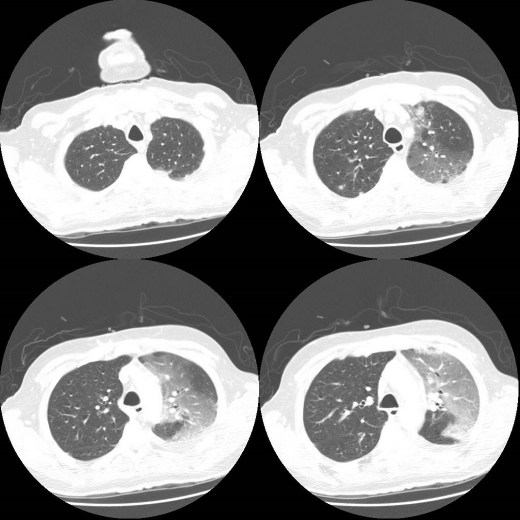
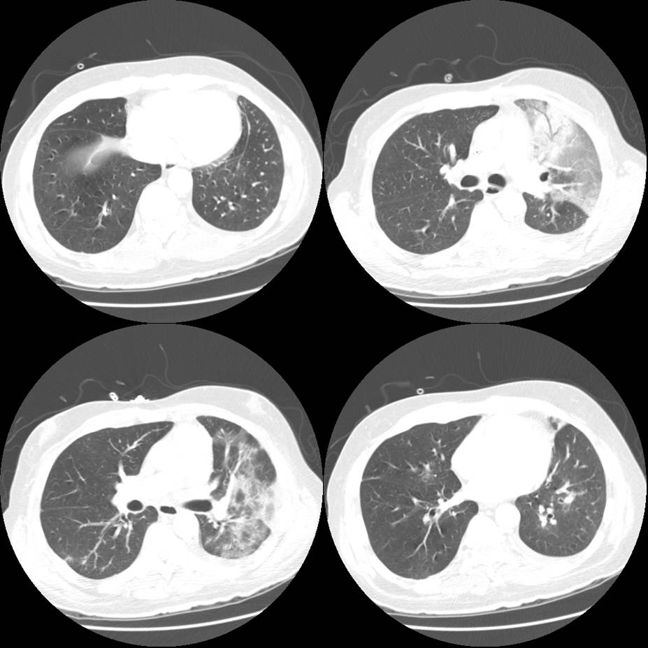
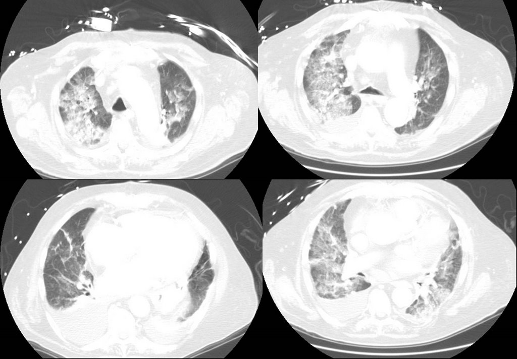
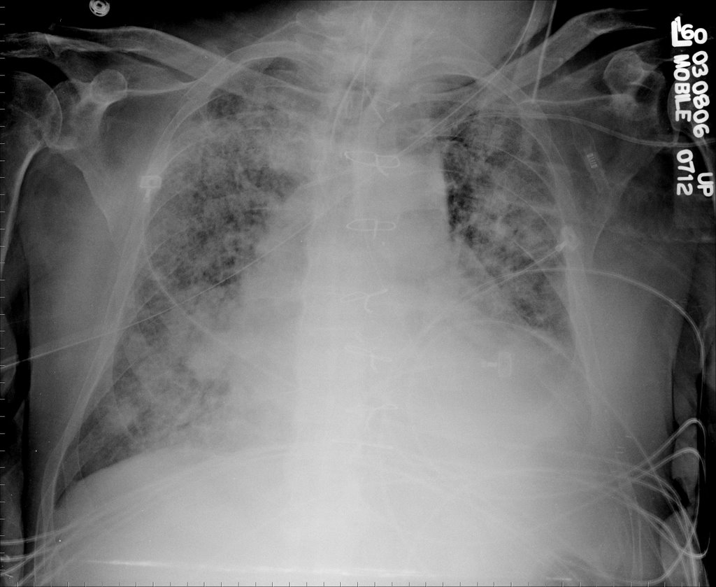
She then developed some respiratory distress, mild hemoptysis, worsening hypoxemia and had to be intubated and had the changes seen on the second CxR. (Some cuts from the CT from that day are included).

What would you want to know/do next?

Monday, September 03, 2007

Abnormal CxR

This is a follow-up to the case below of the 64 y/o man with dense pulmonary infiltrates.
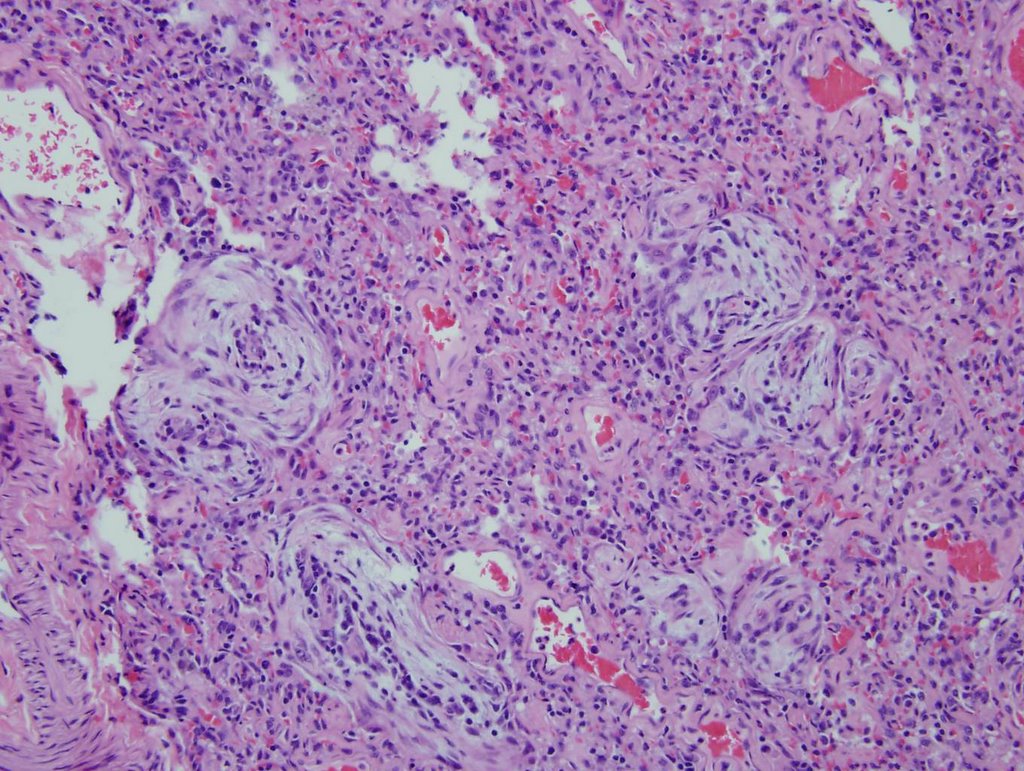
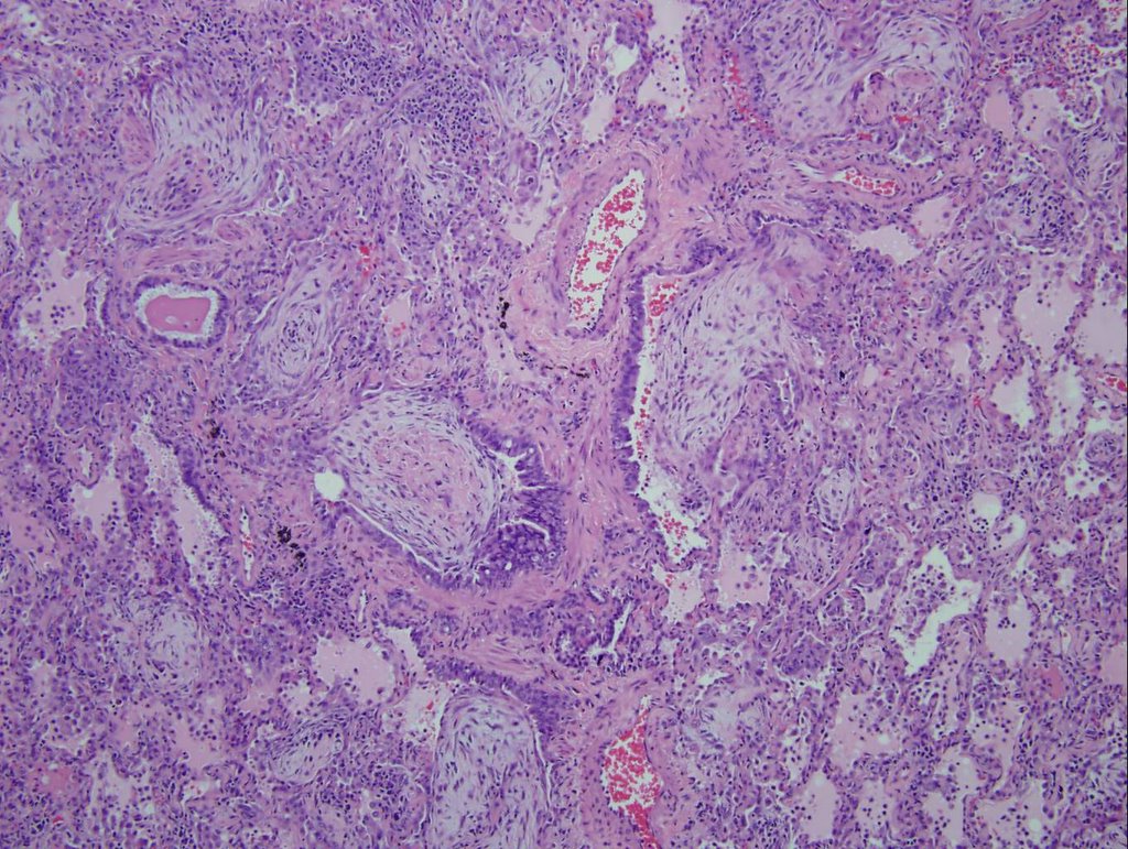
Cultures were all negative. His ANCA was positive with an atypical pattern. A mediastinoscopy revealed large benign nodes with sinus histiocytosis with no diagnostic evidence of metastatic carcinoma, granulomata, or lymphoma.
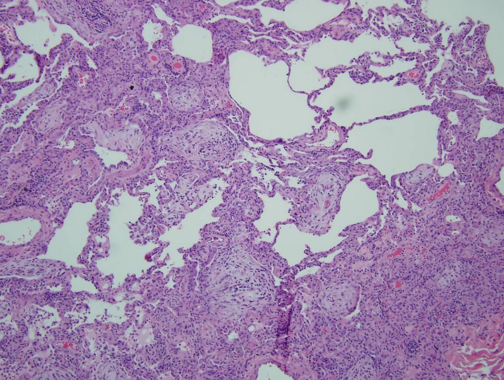
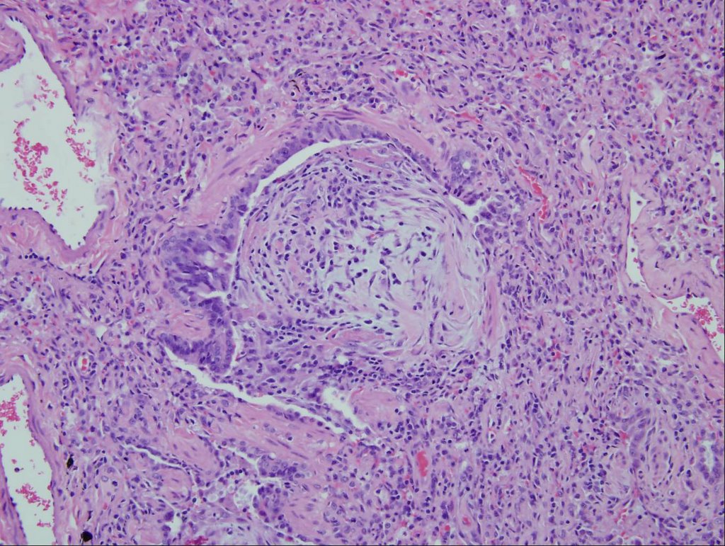
His VATS lung Bx revealed necrotizing vasculitis and fibrosis most consistent with Wegener's granulomatosis (the Bx slides were sent out for a second opinion and were reviewed by Dr. Katzenstein).
How would you treat him?

Friday, April 13, 2007

Cavitary mass

86 year old man with history of dementia brought by his caretaker for mental status changes and lethargy. He lives in an assisted living facility, but has no TB risk factors otherwise (He has his own apartment-like structure, but I beleive there is a common area).

His CT:


There was no hilar or mediastinal adenopathy.
The thickest wall area on that lesion was 4-5 mm.

Someone placed a PPD and it was 11 mm. It was reportedly negative 6 months ago.
He is not productive of sputum, so 3 gastric lavages were done and they were all negative. What would you do next? Are you worried about active tb? Here are my thoughts
1. How sensitive is gastric lavage for afb?
2. location of the cavitary mass is not where tb should be.
3. is he a ppd converter, or was he anergic or are we seeing a booster effect
4. His family does not want an aggressive w/u for cancer, but on the other hand one would not want to send someone home to hospice with active tb....

Tuesday, October 24, 2006

Is this BOOP, Y-J?

41 yo male who works for a satellite dish installation company presented with progressive dyspnea over a couple of months. An urgent care CXR was (+) for a nodule in the R mid lung field, and he was referred to his PCP; he received no intervention at that time. He was lost to f/u and ultimately presented with progressive symptoms one month later. His PCP obtained a CT (see below) and he was sent to be admitted. Upon seeing him, he look chronically ill, but not toxic.


I bronched him, and it was non-diagnostic. I sent him for a surgical biopsy.
The report was read as BOOP with an microscopic sulfur granules c/w actinomycosis.

I started him on Augmentin and saw in the office in 3 weeks.
He was no better. Spiro was c/w were moderate restrictive vent defect.

So, I started him on steroids at 40 mg/day of prednisone x 3 weeks then 30 mg/day.
I saw him 4 weeks later (about 1 week into his 30 mg/day) without any clinical improvement.
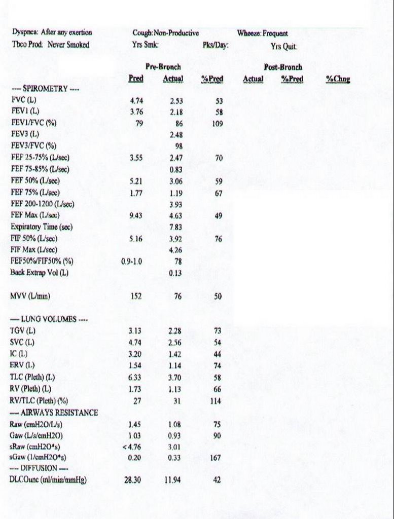
His full PFT's are c/w mild obstruction with air trapping and impaired gas exchange (DL ~70% predicted). He is a smoker, and is still smoking about 1/2 ppd.

I had his biopsy reviewed by Jeff Myers at U of M (profoundly smart and helpful- wish he would have joined the faculty 5 years sooner), but he thought the original interp was correct.

Now what?

Monday, July 24, 2006

More on the woman with eosinophilia

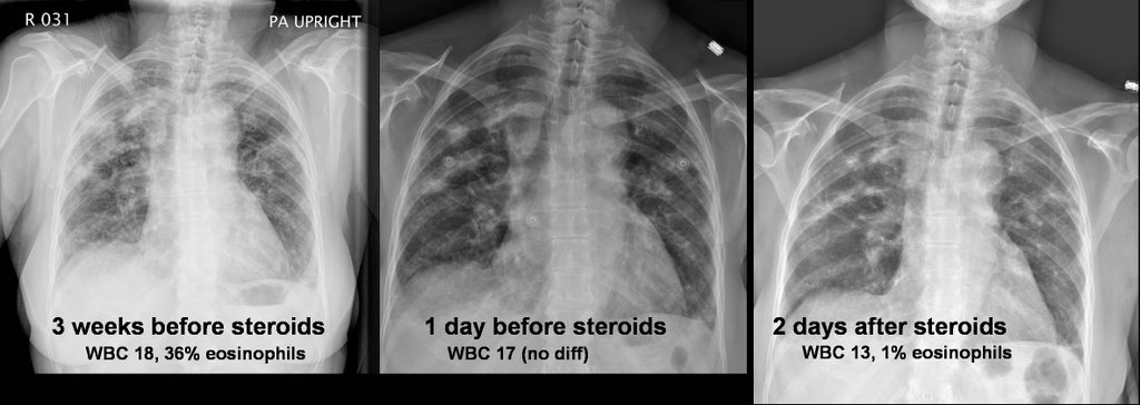
Well, as you might remember, this patient with a past medical history of asthma had had a few months of dyspnea and malaise. A peripheral showed 42% eosinophils and a BAL showed 20% eosinophils. A transbronchial biopsy showed eosinophilic infiltration. Micro was negative. Stool O&P was also negative.
She was given steroids with subjective improvement. However, I am not entirely satisfied with a diagnosis of chronic eosinophilic pneumonia. Here are her radiographs:


Now her amylase and lipase are elevated which does not go with eosinophilic pneumonia, and I don't have a good reason for that. Any thoughts out there? If it is parasitic, at least she is not worsening on the steroids....

Sunday, June 25, 2006

What is causing this RUL process?

54 year old man with AML s/p allogenic bone marrow transplant april 2005 on immunosupp therapy for GVHD of the skin. He is CMV negative by UltraQuant less than 200. He was admitted for shortness of breath and has a RUL infiltrate which you can see below. He was transferred to the ICU because of hypoxemic respiratory failure.

Meds before admission: Prograf 0.5 mg every 12 hours, Medrol 2 mg QOD, Pen VK 500 mg b.i.d., Valtrex 500 mg daily, Diflucan 200 mg daily, pentamidine inhalation every four weeks for PCP prophylaxis

In the ICU, his antibitic coverage was broadened to include tobra/vanco/zosyn/voraconazole and PCP-dose IV bactrim. A BAL showed 250 WBC's with 70% PMNs, but no growth and the gram stain was also negative.


ID consult want an open lung biopsy, but when pressed for what they were looking for were unable to give a specifi entity. What are your thoughts and what would you do next?

Thursday, June 22, 2006

Worsening SOB

This 76 year old man has IPF documented on a 2004 open lung with typical CT findings as well. In 2004 his FVC was 47% predicted with a DLCO of 31% on 6l. It is severe, but he has been stable over a 2 year period. He did have a trial of steroids back then with no effect (no surprise.
He was doing well until a month ago when he developed a "cold" (increased cough with whitish sputum, runny nose/nasal congestion). He continues to have profound DOE. No fevers or chills. 3 separate antibiotic course were without effect. He is SOB even with urination. On exam he can speak in full sentences and is in no acute distress at rest. His sat is 93% on 4l NC. His lungs show no change in his fine bibasilar crackles 1/2 up. JVP about 5-6 cm. No LE swelling. A spiro showed no chnage in mechanics compared to 3 months ago (before the "cold"); FVC 48% pred, but DLCO now is worse at 23% predicted (31% in 2004 the last time it was checked). However, the IVC was only 38% of predicted.
On exam his sat is 93% on 4l at rest.
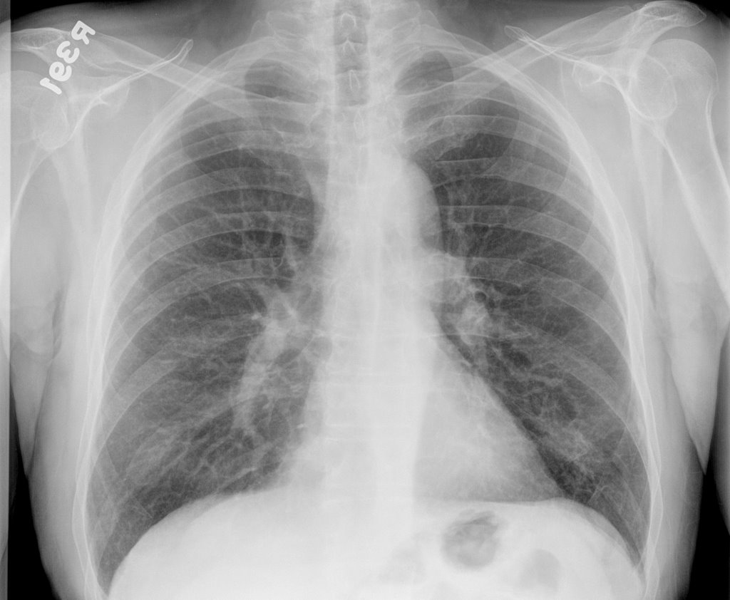
His CXR is unchanged from a year ago and is shown here.


What are your thoughts as to the cause of his new dyspnea? What else would you do?
I just saw him today so there is no "magic" answer that I am witholding; just looking for other opinions (you know, like a roundtable discussion).
Wednesday, May 17, 2006

Follow up to Progressive dyspnea

This is the case of the 71 year old with 3 months of dyspnea. AK astutely pointed to the hot tub and indeed, the patient's use of the hot tub in his new home coincided with the onset of symptoms. A transbronchial biopsy during his illness


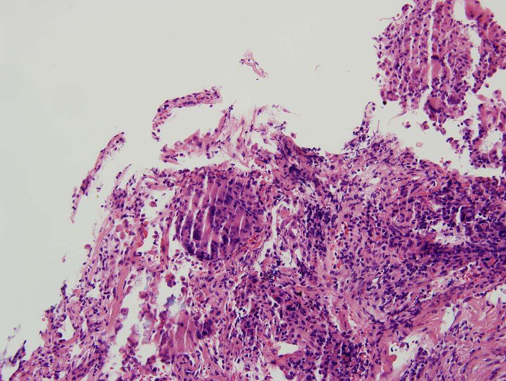
showed chronic and granulomatous inflammation, with focal active lung injury.
The BAL was negative for mycobacteria (specifically mycobacterium avium complex (MAC)). He was started on prednisone 60 mg and his symptoms are essentially resolved after 1 month of therapy. His followup CXR showed clearing of much of the prior acute interstitial infiltrates:


Final Diagnosis: Hypersensitivity pneumonitis ("Hot tub lung")
Monday, May 15, 2006

Progressive dyspnea

71 year old with mild COPD reports progressive dyspnea over the last 2-3 months. He has been treated for his COPD with an Atrovent and an Advair inhaler. He says that his symptoms were relatively well controlled with these medications until recently when his shortness of breath became progressively worse. No cough, fever chills or diaphoresis. Mild weight loss with relative anorexia. He has arthritis and has multiple complaints of joint pains.

PmHx.
1. Hypertension.
2. Benign prostatic hypertrophy.
3. Hypothyroidism.
4. Gastric reflux.
5. Osteoarthritis - What helps is etodolac 400 mg BID and soaking feet in hot tub.
6. COPD.


Meds: Levoxyl, Protonix 40 mg daily, Plendil, terazosin, Atrovent, Advair, and Lodine 400 mg BID, Relpax PRN. For migraine headaches he used to take Cafergot

FHx - no lung or autoimmune disease
Shx - quit smoking 14 years ago after having smoked a pack per day for
35 to 40 years. Lives in a house which is new. He used to live in an apartment a few months ago with no problems there.

Spirometry: FEV1/FVC 68.5 (93%), FEV1 2.85 L (94%)FVC 4.16 L (101%)DLCO 54% predicted.

CPK normal 104, Sed. rate 15, ANA weakly positive, rheumatoid factor negative. He has had normal SPE, aldolase, cortisol, urinalysis, lytes-7 profile, liver profile, TSH, CBC. Cervical spine x-ray showed advanced degenerative changes. X-rays of the hands have shown osteoarthritis mainly at the first carpometacarpal joints. Lumbosacral spine x-ray showed degenerative changes.

Over the course of his progressive dyspnea he had some xray changes.
CXR last year:


During increased dyspnea:

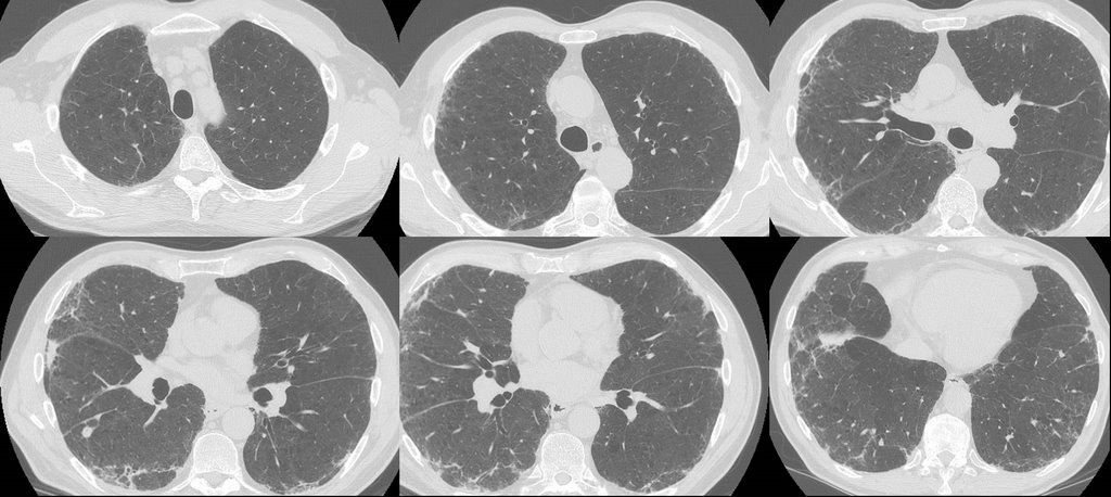
CT around same time shows changes:

What would you do next, or what thoughts do you have regarding the DDx?


Answer is here

Monday, April 24, 2006

Abnormal CT and hypoxia.

38 year old white female with PMH of interstial cystitis presented to ER by EMS after she was found to have a seizure at home witnessed by her mother. That was her second seizure and her initial one was 1 month ago when she went to an outside local hospital. Her work up at that time included head CT which was negative except for sinus disease. She was advised to follow up with her PCP at that time.

In the emergency room, she was afebrile but post-ictal. She was also hypoxic. She was put on oxygen and admitted.
PmHx only significant for the interestitial cystitis. Had some ankle edema 6 years ago treated with low salt diet; thought secondary to her job standing all day (dishwasher).
Medications: none.

CT scan:

RF and sed rate low or normal.

What are your thoughts and what would you do?

Tuesday, April 11, 2006

collapsed RML

Check out Jeff's post below.

I hate to answer a question with another question, Jeff but I have had this similar case. This is a 65 y/o woman, no significant TOB Hx with recurrent partial LLL collapse. She has had LLL CAP 3 times (see above) and though the pneumonias clinically all resolve (they have been over ~ 5years) she always has a residual partial LLL collapse. She was bronch'ed in 2001, 2002 and I have re-bronch'ed her in 2005. Her airway though a little narow with some dynamic collapse looks normal and biopsies and Cxs were also all negative.
Other than encouraging pulmonary clearance, chest P&PD, etc. would you do anything else?

Friday, March 17, 2006

Respiratory failure

60 y/o man PmHx only ITP (controlled with prednisone 40 qd), and hypertension. He had 6 months of SOB, and was found to have severe AI. He underwent valve replacement. His preop CXR is here:
He then developed SIRS (WBC, fever, tachy) but no infectious source ID'd. He was intubated. A CT is here: .
A month later he is still intubated with the following CXR:

What is your differential diagnosis? What tests/procedures/other data would you like to know?

Monday, March 06, 2006

30 year old with progressing infiltrates.

Back in June I posted the case of a 30 year old man no PmHx with 3 months of fevers and malaise. HIV negative. CBC with normal diff without eos. A CT on outpatient is shown here. .


His dyspnea worsened and he was admitted and then intubated. His condition worsened.
For instance, here is a chest xray from one of the ICU days (the sub-cu air is from chest tubes after the VATS.


He did not respoond to steroids and the micro workup was unremarkable. He died after 2 months on the vent.

Here are the biopsies:


I realize there may be no pathologists out there reading this, but what would be your top diagnosis (and I'll post answer later in the day).

Friday, February 24, 2006

Ground glass opacities??

43 yo male with minimal PMH presents with 1 year history of constitutional symptoms (lethargy, intermittent fevers, and RUQ pain. Was diagnosed with elevated transaminases, but a cause (to date) has not been determined.
3-4 weeks ago, became progressively more dyspneic. Initially noted with moderate amts of exertion, but now is at rest. Denies any cough, sputum. Has some pleuritic CP. Still having intermittent fevers, chills and sweats.

No TB risk factors.

PMH: Hypertension, hyperlipidemia
FH: nothing strange (i.e. only HTN, DM2, CAD, etc)

No new meds. He is on a statin, (LFT’s have been stable since placement on this drug 9 months ago), BBlocker, aleve

PE: Temp 38.4; 90% on RA at rest… desaturates to 85% with 4 minutes of walking
Lungs: Clear
Tatoos on both upper extremities (professionally done)

Basic labs normal except AST and ALT in 400’s. They have not appreciably changed in 6 months.
Hepatitis panels negative; Abd u/s shows some “fatter liver” with boarderline splenomegaly

PFT’s


The CXR showed interstitial pattern; to save space, I will not include it. Most would have gotten a CT in f/u anyway.

CT (not high resolution)


What would you do next?

Monday, February 20, 2006

Shortness of breath over a 2 month period

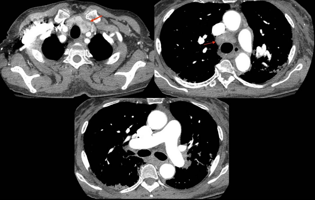
This 50 y/o AA woman presented with shortness of breath. Without further history, what would be your (not necessarily complete) DDX based on the CT. Also note the mediastinal nodes (arrows).