Since another question from a doc is related to TB, I'll post that here as well:
55 y old gentleman presented with few weeks history of progressive dyspne and right sided pleuritic chest pain, with history of contact with a case of pulmonary TB, no symptoms of toxemia, clinically the patient got signs of right sided pleural effusion which was aspirated and shown to be lymphocytic exudate,because the patient also presented with hoaseness of voice CT chest was done showed no lung masses or lymphadenopathy, BAL showed no malignant cells, PPD test was highly positive, laryngeal examination showed cordal polyp. culturing the fluid and sputum for TB was negativethe patient was started on antituberculous ttt , 1st 2 months quadrible therpy and then dual therapy and the patient still have re accumulating effusion? any suggestions?
Showing posts with label Pleural diseases. Show all posts
Showing posts with label Pleural diseases. Show all posts
Friday, November 30, 2007


Monday, September 10, 2007
intermittent oxygen in hydropneumothorax with Bronchopleural fistula(BPF)?
Here is an interesting question submitted by "zolt"
OK we all know that oxygen accelerates the rate of absorption of pneumothorax by about 4 times and so is of value in patients with pneumothorax being managed conservatively. Now in patients with hydropneumothorax with collapse with BPF with tube thoracostomy, is there any role of intermittent oxygen? how will be the diffusion mechanics in such patients or will all the oxygen come out through BPF?
OK we all know that oxygen accelerates the rate of absorption of pneumothorax by about 4 times and so is of value in patients with pneumothorax being managed conservatively. Now in patients with hydropneumothorax with collapse with BPF with tube thoracostomy, is there any role of intermittent oxygen? how will be the diffusion mechanics in such patients or will all the oxygen come out through BPF?
Tuesday, October 10, 2006
Persistent pleural effusion
This is a 38 year old man who is 1.5 years s/p a bone marrow transplant for non-Hodgekin's Lymphoma that had relapsed despite several chemotherapeutic regimens. His post-transplant course has been generally uneventful, with good engraftment, no major infectious complications, and minimal GVHD of the skin and gut. He also had a DVT at some point, treated with adequate anticoagulation for 9 months. He was referred for a right pleural effusion. This was tapped-it was a chylothorax. Flow-cytometry was negative for a recurrence of the lymphoma.
His pulmonary function varies a bit between normal and a mild obstructive ventilatory defect. His exercise capacity has continued to improve since the BMT, but remains significantly below his pre-morbid state (he was, prior to his cancer, a highly trained endurance athlete).
He is referred for pleuradesis.
His films are below:
His pre-BMT film:
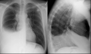
One year later, prior to thoracentesis:
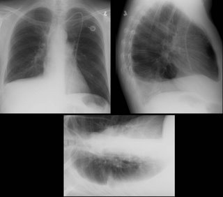
One-day following a 2-liter thoracentesis
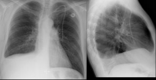
Thoughts? How would you proceed?
His pulmonary function varies a bit between normal and a mild obstructive ventilatory defect. His exercise capacity has continued to improve since the BMT, but remains significantly below his pre-morbid state (he was, prior to his cancer, a highly trained endurance athlete).
He is referred for pleuradesis.
His films are below:
His pre-BMT film:

One year later, prior to thoracentesis:
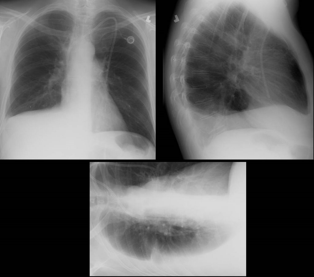
One-day following a 2-liter thoracentesis

Thoughts? How would you proceed?
Monday, September 11, 2006
Bloody effusion
57 y/o man former smoker, presented with pleuritic CP and found to have left loculated effusion. No constitutional sx. no wt loss, fever, chills, cough. Tap 2 weeks later showed it to be bloody, with 57 WBC, 49% PMN and 49% lymphocytes. He is currently asymptomatic.
The effusion was AFB - times 1. The micro and cytology was negatrive. Dopplers of legs negative and V/Q was intermediate or indeterminate (cant remember) and PE-protocol CT was read as negative.
His Pmhx not remarkable except for 2 previous pneumonias. Last was in march and was on the right side. There was a small parapneumonic effusion which has since resolved.
No history of trauma except moving his furniture 1 day before the pleuritic CP, but he did not hit anything per se. No Fmhx of thrmbosis and in fact his Wells criteria is zero.
What would you all do next?
The effusion was AFB - times 1. The micro and cytology was negatrive. Dopplers of legs negative and V/Q was intermediate or indeterminate (cant remember) and PE-protocol CT was read as negative.
His Pmhx not remarkable except for 2 previous pneumonias. Last was in march and was on the right side. There was a small parapneumonic effusion which has since resolved.
No history of trauma except moving his furniture 1 day before the pleuritic CP, but he did not hit anything per se. No Fmhx of thrmbosis and in fact his Wells criteria is zero.
What would you all do next?
Thursday, August 31, 2006
Benign uterine fibroids and pleural effusion
This is an outside submission for a question correlating a pleural effusion and Gyn disease:
Patient with a right pleural effusion, more than 1500 ml tapped. 2nd tap: more than 1700 ml tapped 6 days later. Contrast CT revealed pelvic fibroids.
Effusion results protein, probably assoc. w/ RBC.5 to 11 months prior, sonograms, contrast MRI revealed uterine fibroids 8 to 10 mm in size, stable in size over 6 months. Uterine scrape (not complete D and C) test benign. No indications of ovarian cysts, etc. Hysterectomy not chosen due to stable fibroid size and results of uterine scrape.Heavy coughing for 9 mos. Chronic post-nasal drip for 23 mos.
Allergy tests reveal slight allergy to a few local plants.
Chest xray showed: Pleural effusion right side.
Latest contrast CT pelvic fibroid.Previous ultrasound and contract MRI showed uterine fibroids stable after approx 6 mos.
Effusion samples from 2 taps have no cytology assoc. w/cancer, no pancreatitis, tb/fungus results not yet available. Unremarkable except for protein.
2D echo unremarkable.
Any suggestions on this reaccumulating effusion?
Patient with a right pleural effusion, more than 1500 ml tapped. 2nd tap: more than 1700 ml tapped 6 days later. Contrast CT revealed pelvic fibroids.
Effusion results protein, probably assoc. w/ RBC.5 to 11 months prior, sonograms, contrast MRI revealed uterine fibroids 8 to 10 mm in size, stable in size over 6 months. Uterine scrape (not complete D and C) test benign. No indications of ovarian cysts, etc. Hysterectomy not chosen due to stable fibroid size and results of uterine scrape.Heavy coughing for 9 mos. Chronic post-nasal drip for 23 mos.
Allergy tests reveal slight allergy to a few local plants.
Chest xray showed: Pleural effusion right side.
Latest contrast CT pelvic fibroid.Previous ultrasound and contract MRI showed uterine fibroids stable after approx 6 mos.
Effusion samples from 2 taps have no cytology assoc. w/cancer, no pancreatitis, tb/fungus results not yet available. Unremarkable except for protein.
2D echo unremarkable.
Any suggestions on this reaccumulating effusion?
Monday, July 31, 2006
Follow up to pleuritic pain and fatigue
Old case presented by Mendez is here.
Follow up: Transbronchial bx's revealed multiple epithelioid granulomas. Special stains for AFB and fungi were negative.
Symptoms improved after treatment with oral prednisone which was tapered over several months.
F/U Chest CT (6 months later) revealed interval regression of peribronchovascular nodularity. Previously enlarged hilar/mediastinal lymph nodes no longer enlarged.
Final Dx: Stage II Sarcoid
Follow up: Transbronchial bx's revealed multiple epithelioid granulomas. Special stains for AFB and fungi were negative.
Symptoms improved after treatment with oral prednisone which was tapered over several months.
F/U Chest CT (6 months later) revealed interval regression of peribronchovascular nodularity. Previously enlarged hilar/mediastinal lymph nodes no longer enlarged.
Final Dx: Stage II Sarcoid
Thursday, June 29, 2006
Pneumothorax and pilot's license
This was left as a comment for this case on pneumothorax, but I reposted it here, as it is interesting:
I am a 19-yr old who had a right spontaneous pneumothorax. BP: 130/70-ish Oxygen Sat: 98% HR: 62
Chest tube inserted first, and then VATS done. First chest xray showed my right lung had collapsed to a size of about 10 to 20% of the original size. Subsequent X-rays after the VATS showed that my lung had re-expanded to 90% of its original size. The surgeons warned of a 10% chance of recurrence or occurrence in the other lung.
My question is, I am passionate about flying, and already secured a place in the undergraduate program in Bachelor of Science (Aviation) in Perth's Edith Cowan University, which includes Commercial Pilot's License. They are waiting for my acceptance. Do I still have the medical conditions and requirements to fly?
I am a 19-yr old who had a right spontaneous pneumothorax. BP: 130/70-ish Oxygen Sat: 98% HR: 62
Chest tube inserted first, and then VATS done. First chest xray showed my right lung had collapsed to a size of about 10 to 20% of the original size. Subsequent X-rays after the VATS showed that my lung had re-expanded to 90% of its original size. The surgeons warned of a 10% chance of recurrence or occurrence in the other lung.
My question is, I am passionate about flying, and already secured a place in the undergraduate program in Bachelor of Science (Aviation) in Perth's Edith Cowan University, which includes Commercial Pilot's License. They are waiting for my acceptance. Do I still have the medical conditions and requirements to fly?
Monday, June 19, 2006
Radiology monday
This 72 year old man is asymptomatic and had a routine chest xray as part of a yearly exam from his primary care physician.


Monday, May 01, 2006
Abnormal CT scan
72 year opld with severe COPD developed right sided pleuritic chest pain. No fever chills or increased shortness of breath.
A chest xray at baseline is shown here:
A chest xray at baseline is shown here:
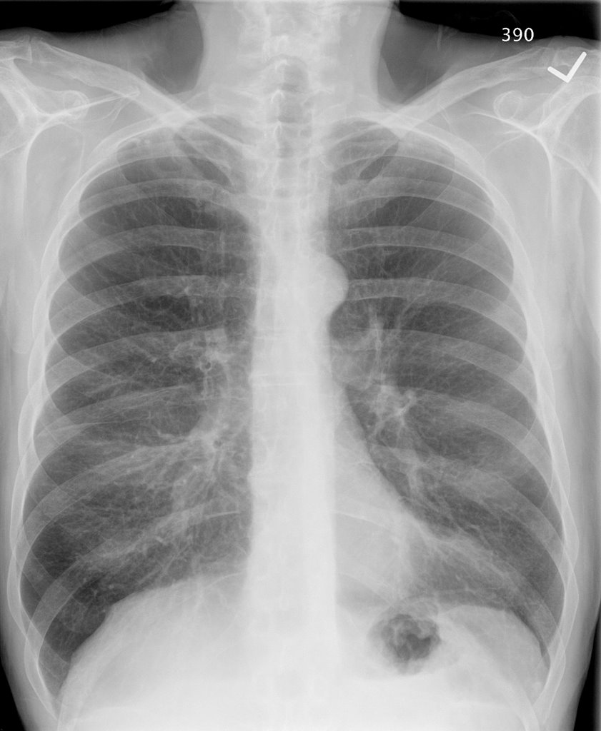
A chest xray with the pain is shown here:

Based on that CXR, he was diagnosed with RUL pneumonia and prescribed a Z-Pak. His pain pertsisted. A month later a f/u chest CT was done. Other than severe upper lobe emphysema, the pertinent slice is shown here:
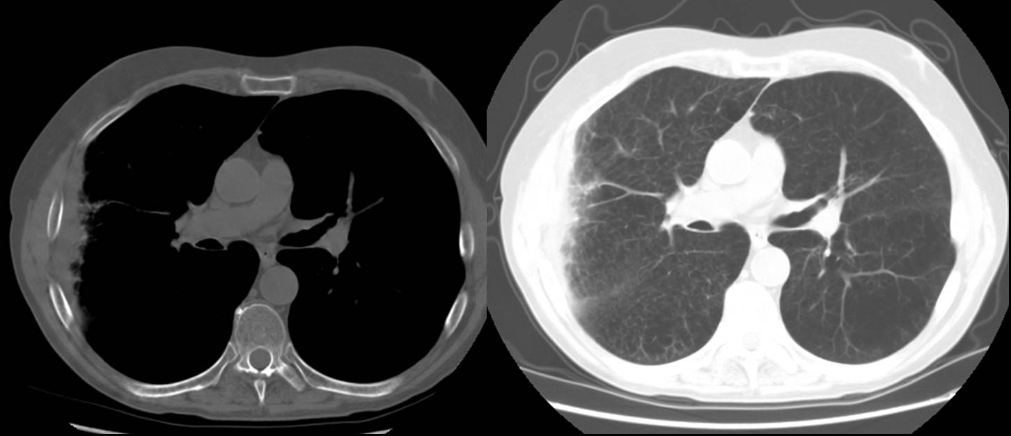
Any comments?
Wednesday, April 05, 2006
PTx

This is just a quick show-and-tell (though feel free to comment).
This is a 73 y/o heavy smoker transferred from an outside hospital with a PTx. He had a CT placed there but had a large persistent leak and was transferred for further evaluation. His outside CT showed lower lobe predominant bullous emphysema with a large RLL bulla. (unfortunately those were outside films and I couldn't post them).
He was started on aggressive bronchodilator therapy and stabilized. His spiro revealed a severe obstructive ventilatory defect. He underwent a bullectomy and the CT surgeon got these great pictures:
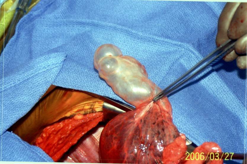
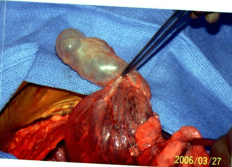
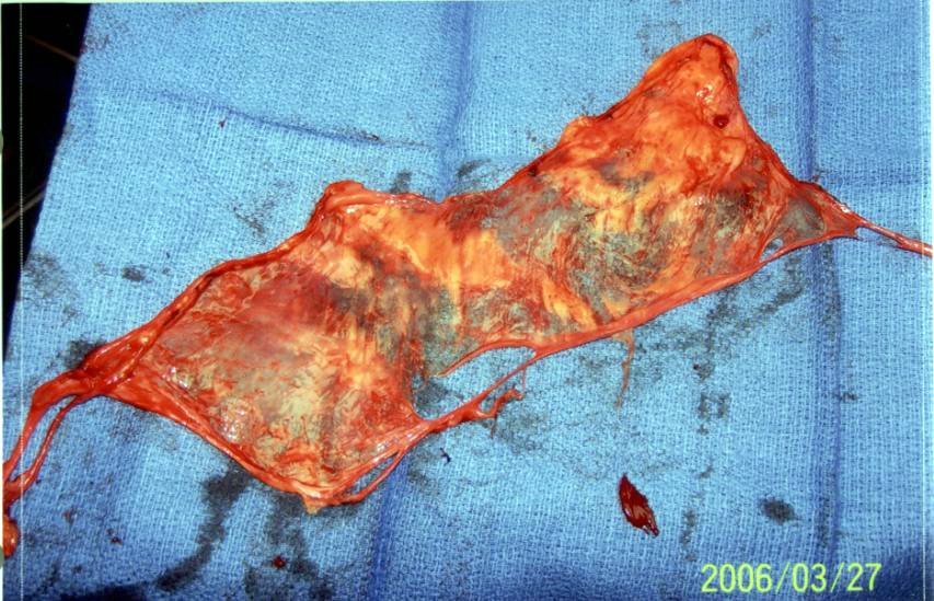
He is doing very well. His BPF has closed with the lobectomy and his dyspnea is much improved.
Thursday, March 30, 2006
Air travel
Dear pulmonologists:
When is it safe to commercial air travel after traumatic hemopneumothorax?
Regards,
Jon Mikel Iñarritu, M.D.
When is it safe to commercial air travel after traumatic hemopneumothorax?
Regards,
Jon Mikel Iñarritu, M.D.
Thursday, March 23, 2006
Follow-up to pneumothorax

This is the young woman with the 30% PTx that was hard to see on the screen. I have played a bit with the contrast... The left film is a repeat from admission with the little red arrows pointing to the lung edge. The one on the right is post placement of a Thoravent (small entry hole, small scar on this young patient) with complete re-expansion. Curiously she had a persistent small leak for a few days but eventually sealed it.
Do you always do a HRCT on this patients? How would you follow her from here?
Tuesday, March 21, 2006
Pneumothorax

I am not sure if the CxR will show up well. This is a healthy, active, non-smoker 18-y/o woman seen by her pediatrician for mild dyspnea and R chest discomfort. She had the CxR on the left with a spontaneous PTx and he decided to wait and follow it up. She had a repeat CxR 4 days later (not shown) with an unchanged PTx. We were called 4 days later by radiology about the persistent PTx on the film on the right. She is very stable. Her Hx is otherwise really unremarkable except for her cousin having had a recurrent PTx requiring a VATS.
What would you do next?
Would you have waited those eight days just "watching" it?
How do you preferentially handle these pneumothoraces (i.e. Cook cath, Thoravent, chest tubes)?
Thursday, March 09, 2006
Follow-up to abnormal CxR
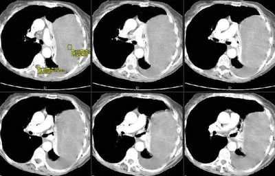
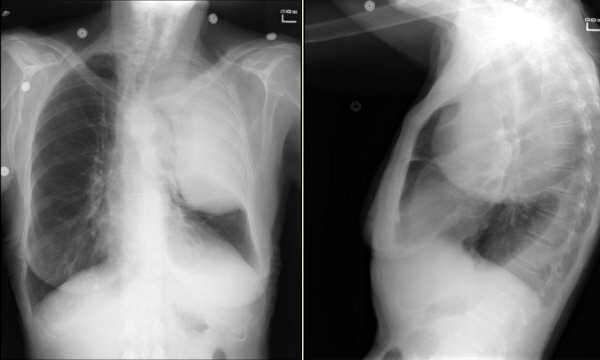
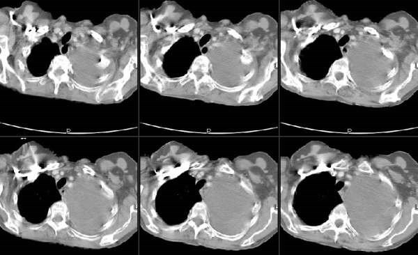
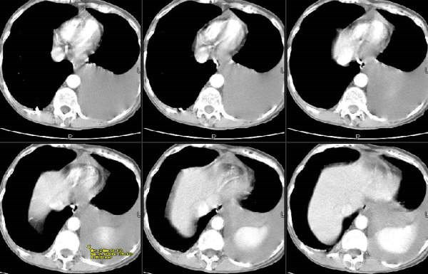
This is the recent post on the 79 y/o woman with a left chest mass. Jeff & Jeff made a good point about the smooth aspect and how the mass looks pleural based. Here are some of the CT images. What do you think?



Thursday, February 16, 2006
Follow up for the chylous effusion
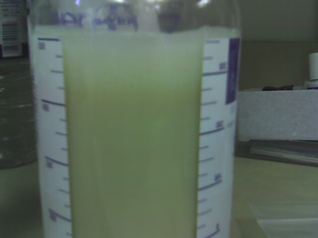
The work-up for this case is ongoing. A colleague of mine is the primary care giver. This patient was referred for an effusion that turned out to be chylous. 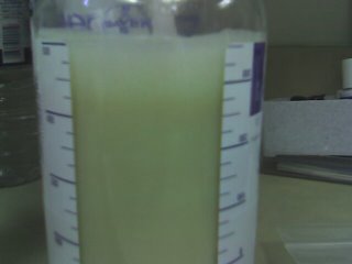
A CT showed a big ol' mass:
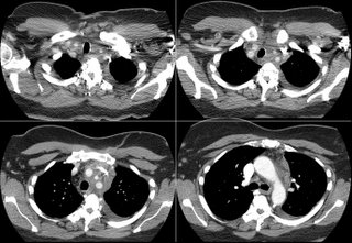 The biopsy is quite interesting. Here's the report: The biopsy contains no normal tissue. All sections contain confluent non-necrotizing granulomas with patchy associated lymphocytic infiltrate. Per the accompanying report, special stains are reported to be negative for fungus and acid fast bacteria.
What are your thoughts about what's going on, and how would you proceed next? There are no "right" answers yet as the workup is ongoing. All comments are helpful.
A CT showed a big ol' mass:
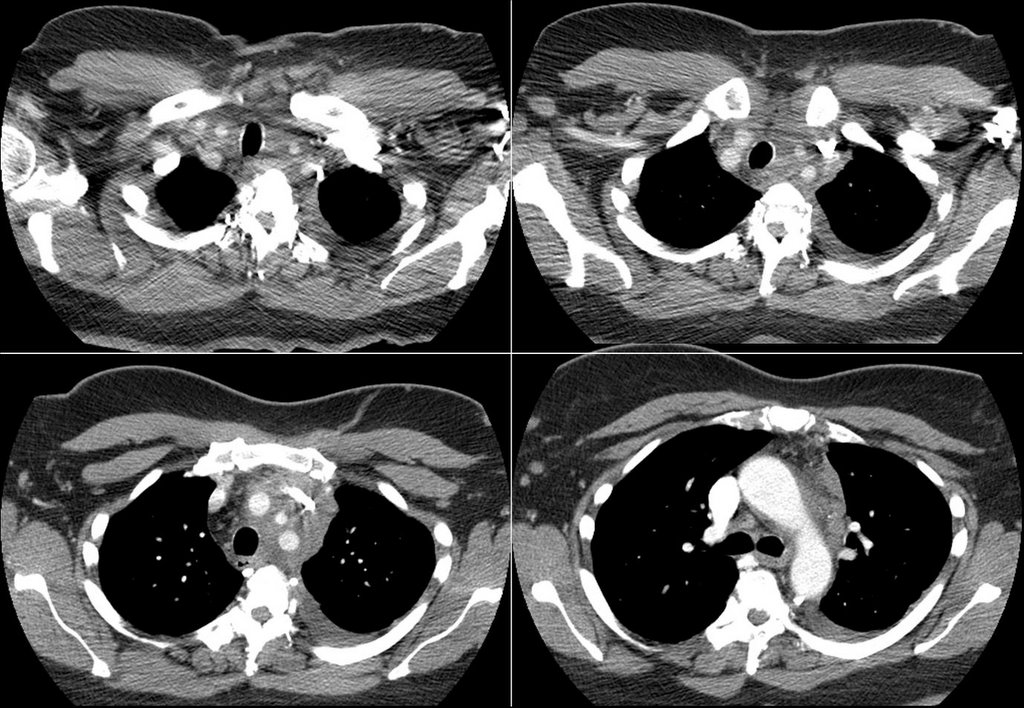 The biopsy is quite interesting. Here's the report: The biopsy contains no normal tissue. All sections contain confluent non-necrotizing granulomas with patchy associated lymphocytic infiltrate. Per the accompanying report, special stains are reported to be negative for fungus and acid fast bacteria.
What are your thoughts about what's going on, and how would you proceed next? There are no "right" answers yet as the workup is ongoing. All comments are helpful.
Subscribe to:
Posts (Atom)