Pt states that was diagnosed with asthma in 2002; at that time she had a CXR that showed scar tissue in the left upper lobe secondary to a previous pneumonia.
She noticed that over the last year, she has being unable to lay down over her right side because she would feel short of breath, so that she always sleep over her left side. Denies frequent cough, sputum production, hemoptysis, weight loss, loss of appetite, chronic chest pain.
SOCIAL HISTORY:
pt has 40 pack year history of smoking, quit in 1998. She works as hair stylist and is exposed to fumes and chemicals. Denies exposure to TB. No recent travels.
PHYSICAL EXAM:
BP 139/83, HR 86x', RR 16x', O2Sat 93% on RA, W 212 pounds.
Patient is awake, alert, obese, oriented x3, no acute distress.
Neck, no lymphadenomegaly. Chest, no use of accesory muscles, no retractions, flat to percussion on the left, no prolonged expiration, absent breath sounds in the left, no wheezes, no crackles. CV: regular rhythm, no S3 or S4, no murmurs. No rubs.
Spirometry showed severe obstruction.
(quick) DDx and how would you proceed?
1 comments - CLICK HERE to read & add your own!:
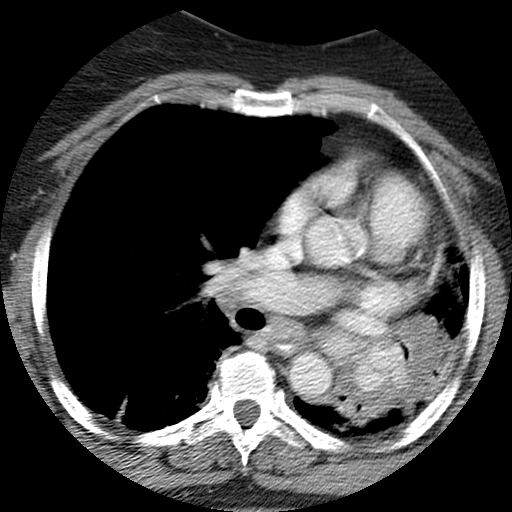
I can't really tell what is going on there on the L. She has marked loos of volume, little lung parenchyma and mixed densities. On that single image I would consider a congenital diaphragmatic hernia with some BPD. Atelectasis/collapse may look like that but without more cuts and lung windows I am not sure why she is so collapsed. I see one cut through a squeezed almost shut bronchus so I guess lung Ca with atelectasis and with the higher density "stuff" pushing against the bronchus, either a broncholith or a hidatid cyst?
Post a Commenttest post a comment