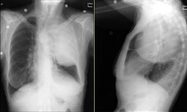
This is the 79 y/o mother of one of our physicians. She has a remote TOB Hx (quit >15 yrs ago) and subacute dyspnea and fatigue (a couple of months'). She presented through the ED with dyspnea and L atypical chest pain. Her nasal swab was positive for influenza and she had the CxRay seen above. What is your DDx and what would you do next?
3 comments - CLICK HERE to read & add your own!:
I *think* the mass is mediastinal. I am not sure if this is superior or anterior division. If superior, lymphoma, thyroid cancer, goiter. If anterior, thymoma. The margins are well-demarcated. This suggests a benign process I would get a CT to sort that out.
I suppose it could also be pleural based; there seems to be some tracheal deviation to the left.
I think it looks pleural based--there is volume loss on the left with mediastinal shift to the right. There's also a smoothly demarcated border extending inferiorly- if that were lobar collapse then the shift should be toward the left.
So, my bet is on a pleural process with a mass-effect shifting the mediastinum to the contralateral side.
I meant tracheal deviation to the *right* when I was talking about the pleural based process...
Post a Commenttest post a comment