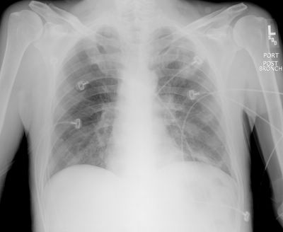
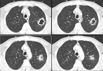
60 y/o male with long smoking history, transferred from OSH for evaluation of an abnormal CxR. He had been admitted to the OSH and failed to respond to IV ABTx (also with a "respiratory" FQ).
He is a limited historian but admits to a persistent cough, purulent sputum production and occasional hemoptysis for "a few weeks". He has lost over 20 lbs in the same period.
PMHx: No label of COPD.
SHx: Quit TOB "a few weeks ago". Used to smoke 2 ppd.
ROS was only remarkable for what was noted above.
On exam, well-nourished, well-developed, overweight male. He has rare occasional ronchi but otherwise clear lungs. Remainder of exam is unremarkable.
Chest radiograph and CT are shown below. What would you do next?


3 comments - CLICK HERE to read & add your own!:
Well, thick walled cavity in a smoker: top on the list is cancer - primary or metastatic. Cannot also exclude TB. Cannot exclude aspergillus. Although the inferior aspect of the cavity is "thicker", I don't see an air-fluid level so not inclined to think of lung abscess. Wegner's can give such a cavity but a single lesion like this would be a bit unusual. I don't think lack of mediastinal adenopathy can rule out anything I put on the differential.
A bronch with BAL sent off for the things listed above would be the next step. I'm not sure what others think, but I would be a little worried about biopsy without confirming that there is no vessel feed (if it's aspergillus), but maybe I am being too cautious?
Once again, Dr. Jennings has beat me to the punch; I agree completely with his post. Top of the list is primary bronchogenic CA--given the cavitation, however, need to consider infectious etiologies. No known immunocomprimise, here, so rare things like Nocardia are not likely but remain in the differential (hey, I have to come up with something that Jeff J didn't allready comment on!).
I agree that a bronch needs to be done. Mediastinal windows aren't shown, so should we assume there are no nodes? We can argue about the potential value of a staging PET, presuming this is carcinoma--either way a bronch with BAL should be done here. I'm not too concerned about doing a biopsy here. Finally, he needs PFT's prior to consideration of any resection.
There no nodes or other cavities hiding elsewhere. I had the same thoughts: he was placed on isolation and a bronch was scheduled.
Post a Commenttest post a comment