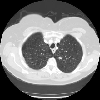
 A CT scan was performed, confirming a 3 cm vertically oriented cylindrical lesion.

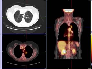
Do you agree that this is an unusual configuration for a malignancy? Could it be a mucous plug? Well, how about the fact that this is the PET scan results:

Discussion of interesting or befuddling cases related to pulmonary and critical care medicine.
 A CT scan was performed, confirming a 3 cm vertically oriented cylindrical lesion.


11 comments - CLICK HERE to read & add your own!:
Is she symptomatic (cough, fevers, etc.)? I may look a little funny-shaped but that PET seems hot. What was the SUV?
I appreciate all the contributors for instructive cases. But I would be very careful not to disclose personal information on imaging studies.
Thank you for the good pick up. The identifiers have been removed. We try to be very compliant with privacy regulations but sometimes things may escape us...
Yes the PET scan is hot, but the cylindrical shape is somewhat atypical for a cancer. It is hard to avoid the possibility that is is an airway plug with inflammation. A bronchoscopy was negative for cancer and the airway exam was unremarkable. The BAL was negative except for 9000 CFU of alpha hemolytic strep.
What would you do now?
Is she symptomatic? It seems a bit far from central airways to have that much inflammation from a plug. Was there a lot of thick secretions in the bronch?
Incidentally, what is her lung function like?
FEV1 is 49% of predicted. At the time of the original CXR showing the 3m lesion, she had URI type symptoms. Currently she just has some dyspnea on exertion but otherwise doing well. As for the bronch there was mildly erythematous airways but without significant secretions. No endobronchial lesions were seen.
You could try some ABTx and bronchodilators and repeat a CT scan in 3 months with close f/up. Was the SUV very high? Though not 100%, at very high levels the likelihood of malignancy vs. infection increases even further...
It is hard to determine the character of the mass from the CT posted. But it seems somehow spiculated. Considering the probability of malignancy from the information given, I would be more inclined to send the pt for VATS. If repeating a CT scan after abx and bronchodilators, I would do it in 6 weeks, not in 3 months.
I guess you could scan him even sooner... if it is inflammatory by 4 weeks we should see a change in the CT. I think the ultimate issue is: do you really believe the PET (I do) and is she in good shape that she would tolerate a curative lobectomy? If yes on both, I agree with Eugene O: send her for VATS with a frozen and extend it into a lobectomy. If she is not a good candidate or if there is any doubt, then 6 weeks or 3 months will make very little difference in the course of the disease and one could repeat a CT then.
How soon to repeat a CT scan in SPN is somehow arbitrary. But a nightmare scenario in this situation is progression to an unrespectable cancer during “ careful watching” like in a well-cited study by O’Rourke N et. al [Clin Oncol 2000;12:141] When it comes to cancer management, it is better safe than sorry especially in light of the current medico-legal climate. I would be very uncomfortable to wait 3 months in this particular case.
I agree that other factors such as operability/resectability, pt’s preference, etc. are very important in the decision-making.
The abnormality does not seem to be in a position that would be ammendable to VATS. She would need an open thoracotomy and her FEV1 is 0.84. The radiologist seems to thing the cylindrical lesion is consistent with a mucous plug. The bronch was negative by cytology and micro and the airway was negative. But I am sensing that the concensus here is to subject the patient to lobectomy?
As for SUV value, our radiologist in nuclear refused to give me the number, saying there was no value in the SUV. He is apparently tracking down articles about the SUV's lack of utility and will get back to me....
Post a Commenttest post a comment