A chest xray at baseline is shown here:
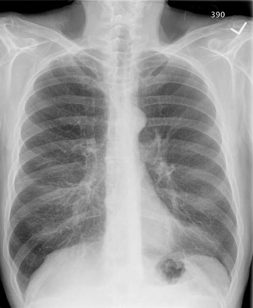
A chest xray with the pain is shown here:

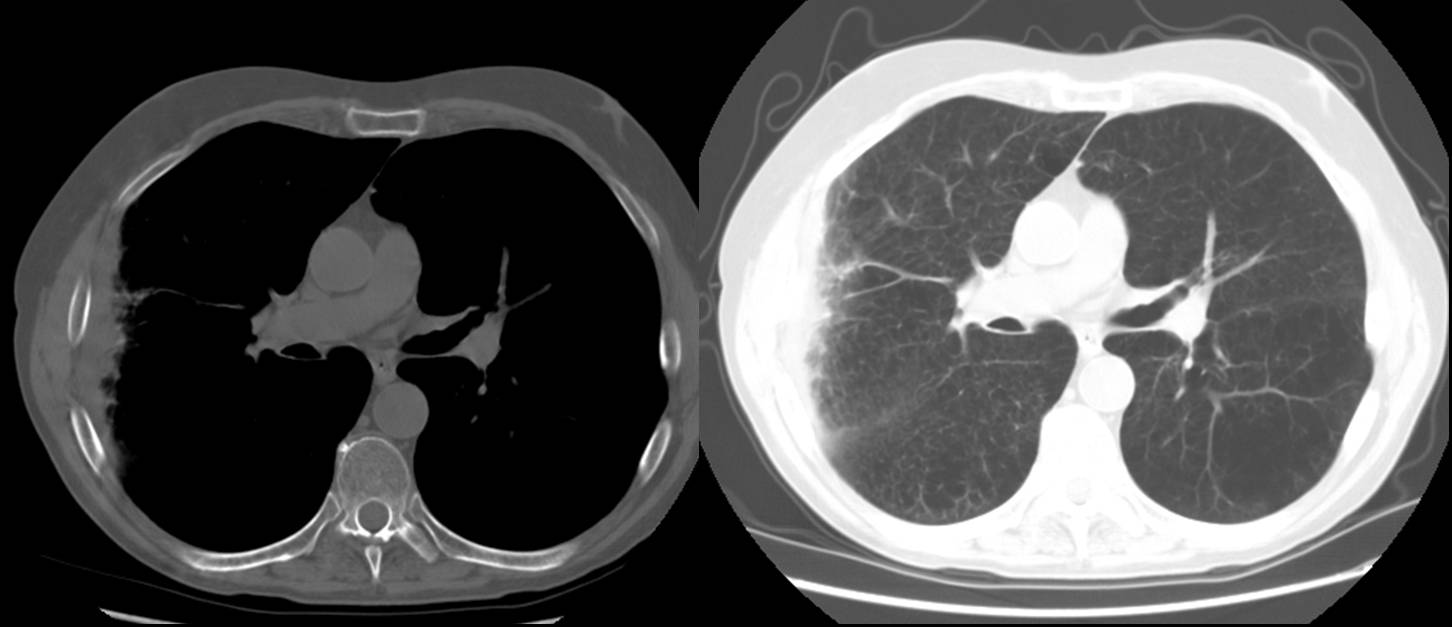
Based on that CXR, he was diagnosed with RUL pneumonia and prescribed a Z-Pak. His pain pertsisted. A month later a f/u chest CT was done. Other than severe upper lobe emphysema, the pertinent slice is shown here:
Any comments?
2 comments - CLICK HERE to read & add your own!:
There is a lot of pleural reaction and a very peripheral infiltrate on the CT. I could not appreciate any plaques on the other films and the pleural-based inflammation seems very focal. COPD with CAP and pleuritis?
Good point and my thought as well. Ironically, this is the one patient that the ER didn't get a PE-protocol CT on. CT has been ordered now, but we are 2 months later and he is asymptomatic so not sure of the yield anymore.
Post a Commenttest post a comment