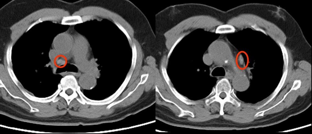
He also has some enlarged nodes (red circles):
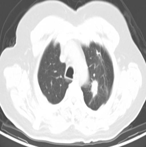
The PET showed intense uptake of the LUL lesion and "mild hypermetabolic activity in the right hilar and pericarinal regions corresponding to nonenlarged lymph nodes on CT. This is of uncertain clinical significance. No other foci of abnormal hypermetabolic activity are identified.":

However, because of the enlarged nodes and the equivocal uptake on PET , I elected to sample them anyway;
The results showed no malignancy from the fine needle aspirate (but there were lymphocytes indicating that the node was sampled.
Not surprisingly, biopsy of the peripheral lesion in the LUL lesion was indeed cancer - adenocarcinoma.
Would you call this cancer a stage I (tumor was 2.4 x 1.4 cm) and proceed to surgery?
3 comments - CLICK HERE to read & add your own!:
I would still be concerned about the nodes. The overall sensitivity of TBNA staging of the mediastinum is 50 percent (of course, with high specificity). And since the larger the lymph node, the greater the likelihood of a positive aspirate and those were fairly small I don't know if you can completely dismiss them. It is clearly the difference between stage I and +N3 dz. I might consider talking it over with the CT surgeons about sampling the nodes.
The nodes are likely benign, in my humble opinion. If they were metastatic, they should be about as hot as the neoplasm in the lung. I live in an area endemic for Histo, and symetric hilar activity is seen on the majority of PETS that I read....and I tend to downplay them.
BTW: Great blog.
I would still probably go with a mediastinoscopy and proceed with lobectomy, if negative. Some aggressive pulmonary rehab pre-operative would fine tune him for surgery and unilung ventilation. A lot of these patients with marginal/ low FEV1's do okay.
Post a Commenttest post a comment