Discussion of interesting or befuddling cases related to pulmonary and critical care medicine.
Tuesday, September 26, 2006
fluid filled lesion
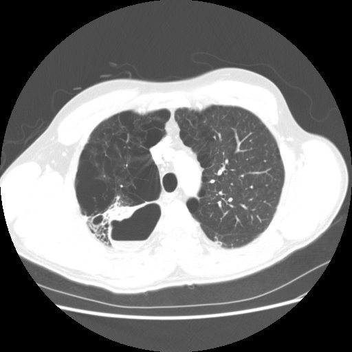
"Anonymous" sent us the following CT slices of a 38 year old who presented with cough and fever. AFB were negative x 3. He was not very productive of sputum and what they did get had many epithelial cells. How would you proceed if this was your patient?
Similar to a case Jennings posted before, I am surprised by the ammount of emphysema in such a young patient... The cavity is fairly thin-walled which carries a much lower RR of melignancy and it is fluid filled. It lloks inflammatory such as a lung abcscess or an infected bulla. I would treat with "typical" ABTx with anaerobic coverage with close radiographic follow-up.
There looks like some associated bronchiectasis, which also supports an infectious/inflammatory etiology. I agree that malignancy is low on the differential. We would need to see more cuts on the CT to ensure that this is not a loculated hydropneumothorax. Presuming it is not, I agree with an anfected bullae, vs. abscess or possibly an infected sequestration. The bronchiectasis may warrant a bronchoscopy to rule out an atypical presentation of mycobacterium.

3 comments - CLICK HERE to read & add your own!:
Similar to a case Jennings posted before, I am surprised by the ammount of emphysema in such a young patient...
The cavity is fairly thin-walled which carries a much lower RR of melignancy and it is fluid filled. It lloks inflammatory such as a lung abcscess or an infected bulla.
I would treat with "typical" ABTx with anaerobic coverage with close radiographic follow-up.
There looks like some associated bronchiectasis, which also supports an infectious/inflammatory etiology. I agree that malignancy is low on the differential. We would need to see more cuts on the CT to ensure that this is not a loculated hydropneumothorax. Presuming it is not, I agree with an anfected bullae, vs. abscess or possibly an infected sequestration. The bronchiectasis may warrant a bronchoscopy to rule out an atypical presentation of mycobacterium.
also consider aspergillosis as a possibility.
Post a Commenttest post a comment