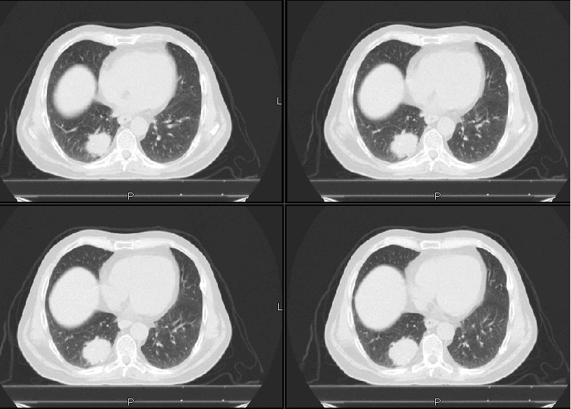
This is a 64 y/o man with a 100-pack-year TOB Hx referred to us for a lung mass found on a CxR for cough. His CT is posted above. The mass is the only abnormality. There is no associated adenopathy.
His FEV1 on no bronchodilators is 1.96L (55%) and he has no other medical problems.
How would you evaluate him? PET, cut, scope?
1 comments - CLICK HERE to read & add your own!:
I am becoming a fan of the staging PET. So, I would start there. If no other lesions are identified, I would proceed with a bronchoscopic airway exam (separately, or in the OR).
Of course, it's allways a bit unnerving to send someone for resection without a tissue diagnosis. But, it looks suspicious and he's got the risk factors, so I I think it's acceptable to go ahead here.
If, however, the PET is negative in that primary lesion, I would still favor resection but would not argue much with short term (6 week or so) reimaging or an attempted needle biopsy.
Post a Commenttest post a comment