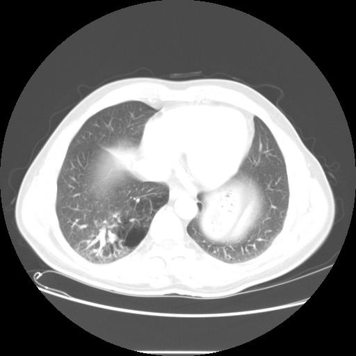
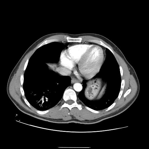
His CT is shown here:
 _______
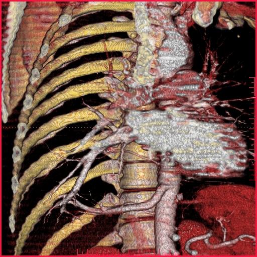
Here is the CT reconstruction:
What is the abnormality and what's your differential. Dont worry about guessing way off base, just guess!
Discussion of interesting or befuddling cases related to pulmonary and critical care medicine.
_______
2 comments - CLICK HERE to read & add your own!:
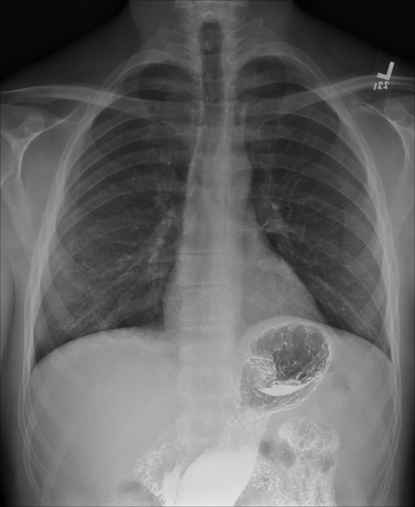
The abnormality on the CT and the CxR looks more like a hollow viscus (e.g. small bowel) in the thorax. The extra vessel seems to originate from the aorta almost below the diaphragm. It seems like a congenital hernia or a congenital cystic adenomatoid malformation (which will be more "hollow" than bronchopulmonary sequestration) rather than a lung sequestration...
See follow-up above. Yeah this reconstruciton is cool because you can see the arterial supply AND the venous drainage back to the atrium.
Post a Commenttest post a comment