35 yo female post-partum x 7 weeks (healthy newborn girl; no complications with pregnancy or with birth) with no PMH presented with the sensation of food getting caught in her throat after eating a large steak meal.
EGD was performed with foodstuff visualized in the proximal esophagus. It was pushed successfully into the stomach.
Post-procedure, the patient had onset of fever (38.5) on one occassion and mild chest pain that resolved without intervention.
Her WBC count was normal and did not change throughout her hospital course.
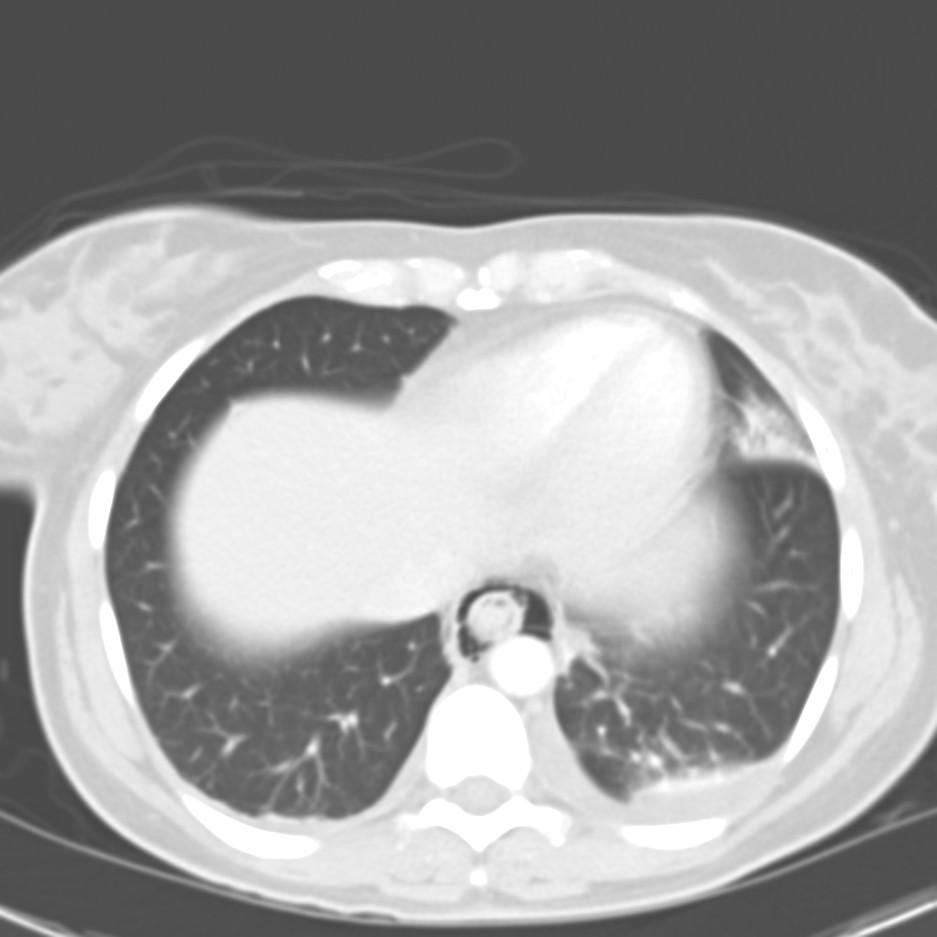
A CT chest was obtained to evaluate.

How would you proceed?
Answer below with discussion.
10 comments - CLICK HERE to read & add your own!:
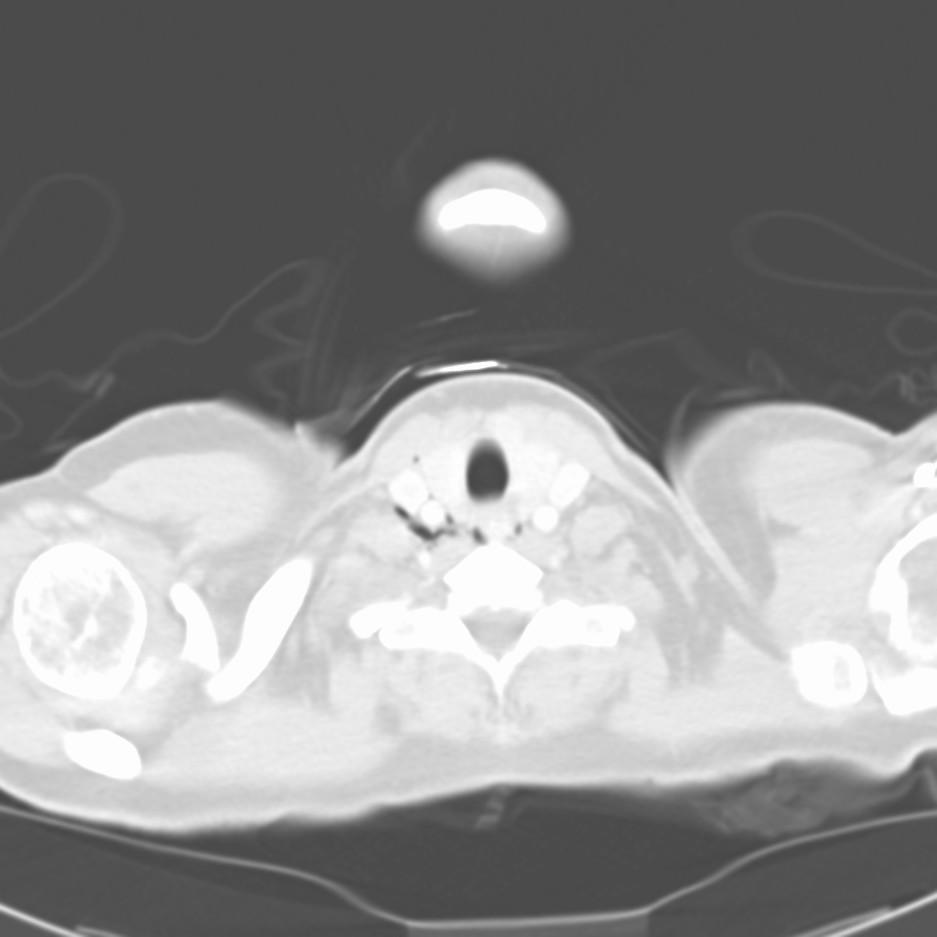
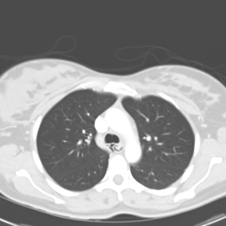
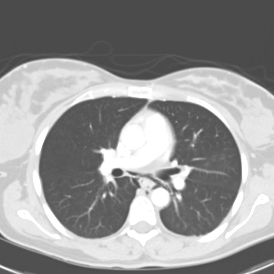
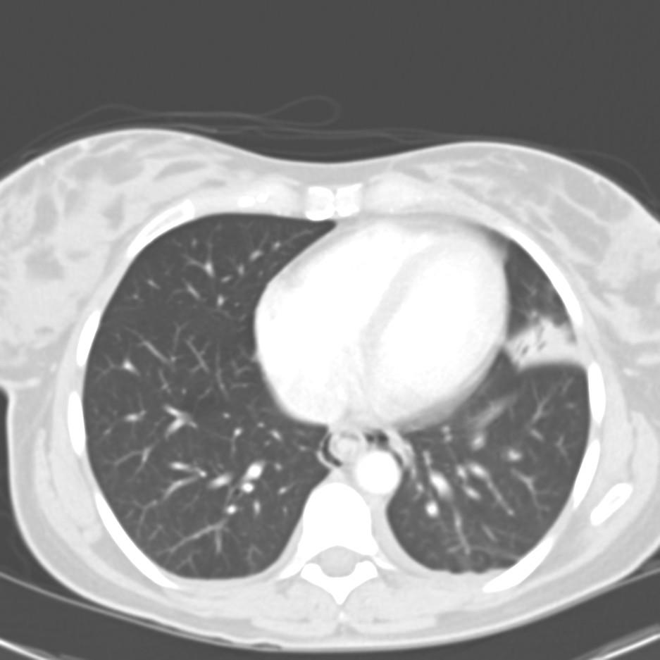
Yea, I agree. There is posterior mediastinal air circumferentially around the esophagus. Looks like a lingular infiltrate/opacity as well and a left pleural effusion.
On top of all that, it looks like there is still a lot of food (or something) throughtout the esophageal lumen. I'd be very worried here.
Yep - looks like the classic pneumoesophogus. One of the known complictions of an EGD - especially since they were mucking around trying to push the food back into the esophagus. HOWEVER, it also can be associated with (bad) GERD - there is the very small chance that she had the air as a result of the original episode of food getting stuck. Only a pre-EGD CT scan would prove this...
These were my thoughts when I first saw the CT (before rounds).
When I looked at her, she is non-toxic, talking on the phone, hungry and upset because she really wants to be at home with her newborn.
The infiltrate and pleural effusion resolved on CXR on hospital day 2. She had a negative esophagram for extravasation of contrast or any other abnormalities.
Now what?
Well, now I have no idea. I've never heard of an esophageal dissection (i.e. mucosal tear with the introduction of air but containment be the adventitia.) I suppose if that could happen, and the mucosal tear was small it might close and the air would be reabsorbed.
Hard to imagine an early tracheo-esophageal fistula forming...
Otherwise, I'm at a loss. I'd ask my gastroenterology colleagues for an explanation!
I'm with Jeff H as well: consult GI. I am surprised at how well she appears to you. With the air and a L effusion I would think (as everyone else did) of an esophageal rupture but it just doesn't fit the picture...
GI was not very helpful.
Thoracic surgery was consulted, and felt that this was likely a common problem that occured because of esophageal manipulation as the esophagus does not have a serosa protecting it.
The T-surgeon mentioned that the GI docs typically use air in the scope to prevent collapsing of the lumen when doing an endoscopy; this is the source of the pneumomediastinum. There was probably had a small tear in the esophagus that closed by the time of the esophagram.
With the progesterone induced weakness (i.e. from pregnancy) in her musculature, I suppose her risk would be higher then the typical young women.
The T-surgeon recommended antibiotics x 2 weeks and 5 days of NPO. If she, on a repeat esophagram, showed no evidence of leaking, a clear liquid diet for 2 weeks and full diet thereafter.
UpToDate has a nice section on this :
http://www.utdol.com/application/topic.asp?file=esophdis/9394&type=A&selectedTitle=3~17
So, your mother was right... chew your food completely before swallowing.
Rather interesting blog you've got here. Thanks the author for it. I like such topics and anything connected to this matter. BTW, why don't you change design :).
I am desperate for help and no one seems to have the answers. I just recently has a CT of the chest and neck done due to dyspnea and chest pain (mostly on the left side by my hear). They were no major findings other than a tiny Pneumomediastinum in the middle medistinum, from the level of the carina to base of the neck. No sighn of mediastinitis or fluids were found. They told me that it should dissolve on it's own after a little while. It has been 3 weeks since the CT and I KNOW that it has not dissolved itself for I can literally FEEL IT! The shortness of breath and the chest pains are not getting any better either. I don't even know how this could have happened. The pulmonologist asked me about any recent injuries. The only thing is that I could think of was an endoscopy I had done but that was over 6 months ago. Other than that, I am a dancer/skater and I work out a lot. I can't think of anything else. PLEASE help me with a piece of advice...anything! NOBODY seems to know anything. I am slowly but surely losing all my faith in all doctors :(
I went from training for the Olympics to not being able to breathe.
If you have any information, you can contact me on here or at:
veronajianni@yahoo.com
Thank you!
Post a Commenttest post a comment