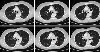
This 49 y/o woman was referred to me with an anormal CT scan. She has a 30 pack/year Hx and has quit smoking when told about the CT. She has no symptoms (she had had a "screening" CxR which prompted the CT), her exam is quite unremarkable and she has normal PFTs (FEV1 and FVC are >85% though the ratio is a bit low).
Her CT showed multiple non-calcified nodules (some a depicted below) with no adenopathy.
What would you do next?
Subscribe to:
Post Comments (Atom)

2 comments - CLICK HERE to read & add your own!:
I'll start with this: the likelihood of lung cancer in a 40 year old, asymptomatic woman is low to begin with. Then, we add that she has bilateral nodules-so that if these are lung cancer, she has either Stage 4 metastatic disease or synchronous primaries. Each is possible, but unlikely.
Could these be metastatic? I'd consider ovarian, less likley breast, possibly germ-cell, lymphoma. Again, I think these are unlikely.
Infectious causes? Possibly endemic fungi and they just havn't calcified yet. Without any symptoms, I think that infections/inflammatory lesions are unlikely.
AVM? Hamartoma? These don't have the classic look.
Autoimmune (i.e. Wegeners)-again, seem less likely without any associated symptoms.
All that said, these aren't the 3mm ditzels so commonly seen today. I think that, as I'm favor a benign lesion here, I'd start with a PET and some screening serologies (ANCA). If all are negative, I'd follow radiagraphically. If the PET is positive, it would get more complicated and depend on whether one or both of these are hot and if there is anything that is extra-thoracic.
I love the fact that we are clustering cases. It really solidifies my understanding of a process if I see several cases with both subtle and not-so-subtle differences.
JCH's differential is excellent.
1. How large are these nodules (i.e. will a negative PET be sensitive enough for us).
2. If DA reads this case, how di you proceed in these cases before the PET scan?
Post a Commenttest post a comment