ROS: Some minor arthralgias, probably OA; o/w unremarkable.
PmHx: chronic UTI's followed by urology otherwise negative.
SH: Lifelong nonsmoker. Occupation was secretary.
FH: 2 daughters with fibromyalgia.
Travel Hx: Thailand in the past year.
On exam initially the lungs were clear to auscultation, but subsequent exams over the next year revelaed bilateral crackles, as can be imagined based on xrays shown below.
So over the next year she became more and more short of breath with the following progression on chest xray.
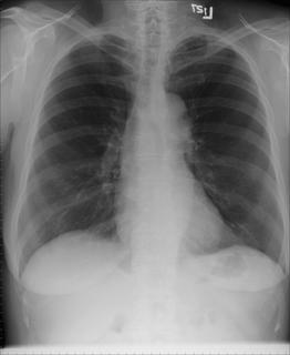 First CXR
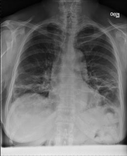
CXR a few months later
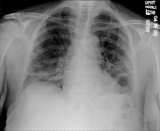
CXR more months later with worsening dyspnea.
Here is a few CT slices around the time of the second chest xray (above):
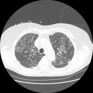
ct up
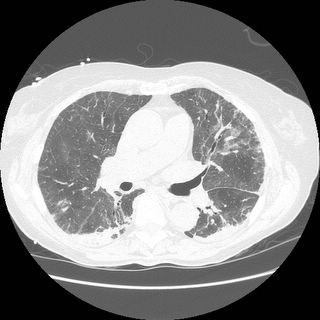
ct mid

ctlow
What additional information would you like to know?
RF and ANA were low in the past.
3 comments - CLICK HERE to read & add your own!:
What do her PFTs look like?
We have 2 PFT's both are when she was already SOB deparated by a good many months. They both show the same mechanics: FEV1/FVC 70 (94%) FEV1 2.43 (100%) FVC 3.45 (107) but the second one also had a DLCO of 56%. They never checked a TLC, but I'm sure you feel it is pretty obviously a restriction.
I think we need to get a bronch and a biopsy. The differential diagnosis of ground glass opacities in an elderly woman is pretty vast including ILD, BOOP, HP, atypical infection, etc., etc.
Please give us the differential of the BAL.
Ultimately, I think you may need to get a SLB to get an answer.
Why did she not get a TLC?
Post a Commenttest post a comment