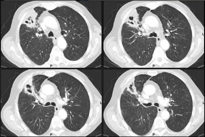
This is a 68 y/o man with COPD, still smoking with a persistent cough. No hemoptysis, no sputum production. His Cxr at his PCP's was abnormal and he came to us with the following CT scan.
SHx:>50 p/year TOB.
Exam showed only hyperinflation and minimal crackles in the R mid lung field.
FVC is 70% and FEV1 is 54%.
What is your differential and how would you proceed from here?
Subscribe to:
Post Comments (Atom)

5 comments - CLICK HERE to read & add your own!:
1. cancer with or without post obstructive pneumonia. This would be any of the NSCLC's, but squamous would go a bit higher.
2. Im not sure if thats a cavity or bronchiectasis. Either way, I would wonder about MAC or another atypical mycobacteria.
3. It looks close to the pleura; I don't know if there is pleural involvement in which case things like actinomycetes might be considered (especially with poor dentition)
4. Speaking of which, an aspiration event into the rml with ensuing chronic smoldering pneumonia might be a possibility.
I would bronch to do airway for an endobronchil lesion then BAL for cytolology, and micro and afb etc.
I agree with jeff (especially with the thought of cancer with post obstructive pneumonia) with the following exceptions:
1. why not regular TB? upper lobe disease with cavities...
2. how about "plain ol' pneumonia" with cavitation(staph,klebsiella,...)
3. I don't think that this is the middle lobe. It is the upper lobe.
I would get sputum samples for gram stain/cultures and AFB, sputum cytology and treat with antibiotics, including anaerobic coverage pending results. also place PPD. If preliminary results negative and there is no clinical/radiographic improvement then Bronch/TBBX/BAL
JJ is actually right, that is the RML. It is more obvious with the other cuts (sorry). There was no adenopathy and the images include the only abnormality. I will give the other bloggers a chance to add their thoughts and include more info/follow-up tomorrow.
Jennings differential is pretty exhaustive.
However, RML syndrome was not specifically outlined.
Anything on the mediastinal cuts?
Anything in the liver/adrenals?
Nothing on mediastinum...
I actually did a bronch on him this past week. Lots of changes of chronic bronchitis. No endobronchial lesions. RML and RUL looked very normal... TBBx unfortunately was non-diagnostic (?): lots of non-specific inflammation and negative smears, with negative Cxs so far.
He feels much better after some steroids and ABTx so we will repeat films in a few weeks and I will keep you posted.
He, of course, continues to smoke a pack-a-day.
Post a Commenttest post a comment