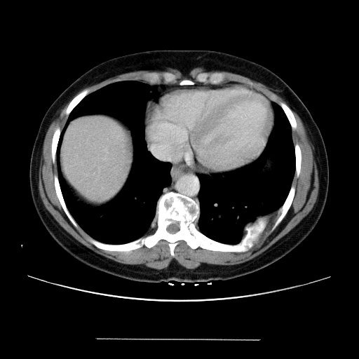
Is that the rib? There seems to be a lot of marrow expansion on that rib mass. Any previous trauma? Without much other Hx, I'd suggest metastatic carcinoma, plasmacytoma (or limited MM) and less likely a primary bone tumor (osteosarc vs osteoid osteoma). Since you selected those cuts, I would guess there was no other obvious mass?
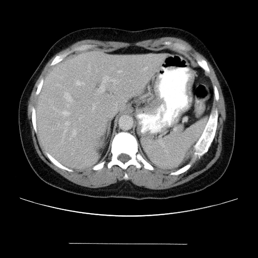
The rest of the CT was normal. She had absolutely no symptoms excep for back pain (which led to the CT). She has no history of any trauma. Oh and in terms of thre pain, there are no neurological deficits of any kind. All of her labs are basically normal. Urine is unremarkable as well.
It turns out that this is fibrous dysplasia, which is a, chronic disorder in which bone expands due to abnormal development of fibrous tissue. There is a defect in osteoblastic differentiation and maturation. This can be diagnosed just on radiograph (shown in slices I provided). You do not need a biopsy unless there is ambiguity in the radiograph. Clues that go against the metastatic bone cancer is that the cortex is intact. Of course, this condition can result in pain and brittle bones, so it's not exactly benign. Her back pain may have been a red herring.
For more info see http://www.emedicine.com/RADIO/topic284.htm

4 comments - CLICK HERE to read & add your own!:
Is that the rib? There seems to be a lot of marrow expansion on that rib mass. Any previous trauma?
Without much other Hx, I'd suggest metastatic carcinoma, plasmacytoma (or limited MM) and less likely a primary bone tumor (osteosarc vs osteoid osteoma).
Since you selected those cuts, I would guess there was no other obvious mass?
The rib is also pretty deformed on that first cut. Any previous trauma?
The rest of the CT was normal. She had absolutely no symptoms excep for back pain (which led to the CT). She has no history of any trauma. Oh and in terms of thre pain, there are no neurological deficits of any kind.
All of her labs are basically normal. Urine is unremarkable as well.
It turns out that this is fibrous dysplasia, which is a, chronic disorder in which bone expands due to abnormal development of fibrous tissue. There is a defect in osteoblastic differentiation and maturation. This can be diagnosed just on radiograph (shown in slices I provided). You do not need a biopsy unless there is ambiguity in the radiograph. Clues that go against the metastatic bone cancer is that the cortex is intact. Of course, this condition can result in pain and brittle bones, so it's not exactly benign.
Her back pain may have been a red herring.
For more info see http://www.emedicine.com/RADIO/topic284.htm
Post a Commenttest post a comment