Wednesday, August 31, 2005
Follow-up to the patient with a solitary lesion
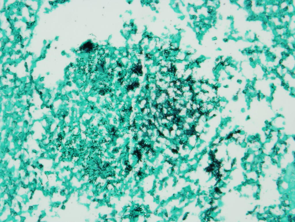
The cavitary lesion was removed. Here is the specimen: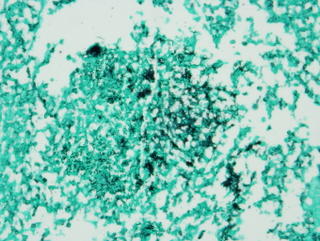
Subscribe to:
Post Comments (Atom)
Discussion of interesting or befuddling cases related to pulmonary and critical care medicine.
5 comments - CLICK HERE to read & add your own!:
oooookay. My pathology is poor, and my microbe identification is worse, but I see some conglomerates of black squiggly-looking things. Looks fungoid, but I can't make out much detail regarding branching (45 degrees vs. 90 degrees.) So, I'm thinking Aspergillus or Mucor, although I'm guessing this can still be Actinomycetes. This is when I go down to the lab, or to my friendly-neighborhood microbiologist, or ID...
I agree that it kind of looks like the whole 45 vs 90 degree thing we all love to talk about. However, the correct bug is what you said third; namely, this is actinomycetes. The official interpretation by our pathologist: right middle lobe contains a cavitary space.The cavity contains
inflammatory exudate and, in one section, a filamentous bacteria
consistent with Actinomycete (gram positive, Grocott stain positive,
Ziehl-Neelsen stain negative). The surrounding lung tissue has a marked
bronchocentric/ bronchiolocentric chronic inflammatory cell infiltrate,
BOOP-like injury pattern, and small granulomas; there is also extensive
perivascular cuffing, follicular bronchitis, and patchy acute
bronchopneumonia.
Very cool. Lately I have been seeing many patients with abnormal TBBx and some wedges in which the pathologists find organisms that look like actino but with no other clinical picture of actino and no + Cxs. In many of these the pathologist can't differentiate oral contamination of the samples from true disease. The Bx in your case looks pretty impressive with the bug at the core of the lesion.
Did you do any more Cxs? Any more ABTx?
Remember, this tissue is from an open lung biopsy where they took the whole lesion out en masse. Therefore, contamination is less likely.
The patient was treated with 6 weeks of pen-G and did fine. He re-presented about 2 weeks post-op with some hemoptysis (airway exam negative though) and an effusion that was bloody. I am assuming this was a surgical complication; all cultures of the effusion were negative for micro. Sorry I dont know the hematocrit of the effusion. My rottion ended and I haven't followed up with the rest of the hospital course; I'll post an update if they get an answer.
Post a Commenttest post a comment