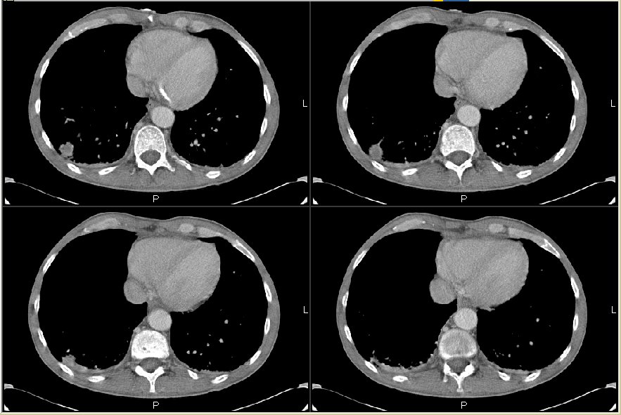
His Cxr showed some hyperinflation and a RLL pulmonary nodule better characterized on the CT scan below (check out the vertebral bodies).
What would you do next? What is your differential?
Answer/follow-up to this case on http://pulmonaryroundtable.blogspot.com/2005/08/follow-up-to-hypercalcemia.html
3 comments - CLICK HERE to read & add your own!:
It looks like a plasmacytoma involving the vertebral body. The lesion on the right side looks like it may also be plasmacytoma and projecting into the thoracic cavity. The only problem with that diagnosis is that you don't usually get hypercalcemia.
Therefore, maybe this is a lung cancer with bony mets. You need a tissue dx and ultimately confirmation that the bone lesion represents cancer. I wonder if a biopsy under guidance would be the best bet so you can diagnose and stage at the same time.
Just curious - which test would be applicable here (in terms of that bone lesion); a PET or a bone scan?
I agree with Jennings; a tissue diagnosis is essential. As you did not show any mediastinal cuts, I'm assuming there are no enlarged nodes.
The nodule itself is relatively circumferential, but appears to have a necrotic core. Additionally, the pleura on the right looks thick and irregular.
I actually think that I would try to get a CT-guided biopsy of the bone lesion first. As to PET vs. bone scan, I think that both should be positive here, presuming that the bone lesion is a met and that the tumor itself is PET +.
If the bone lesion is negative (which I would doubt), then I'd do a staging PET and, if negative, resect the nodule (regardless of the histology).
Post a Commenttest post a comment