Tuesday, November 29, 2005

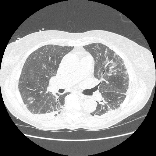
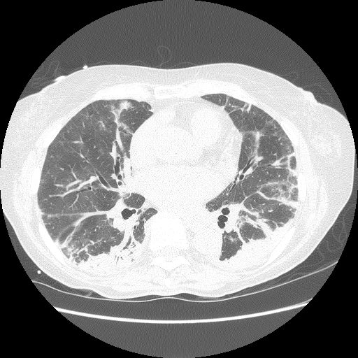
Abnormal CxR and hypoxemia

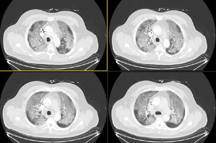
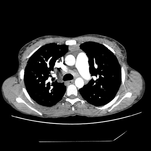
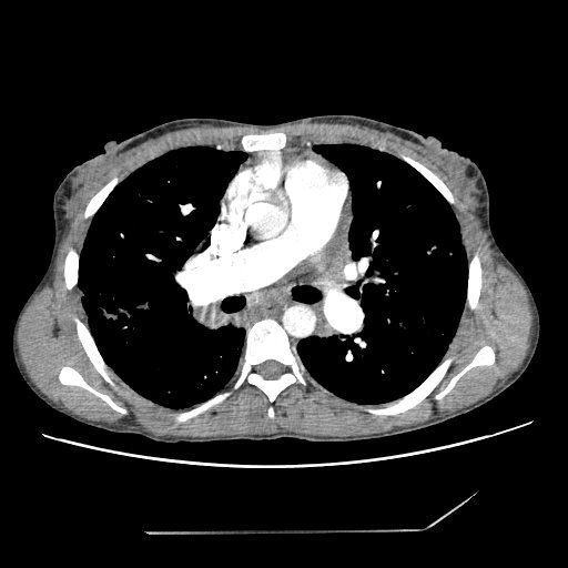
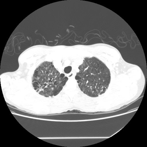
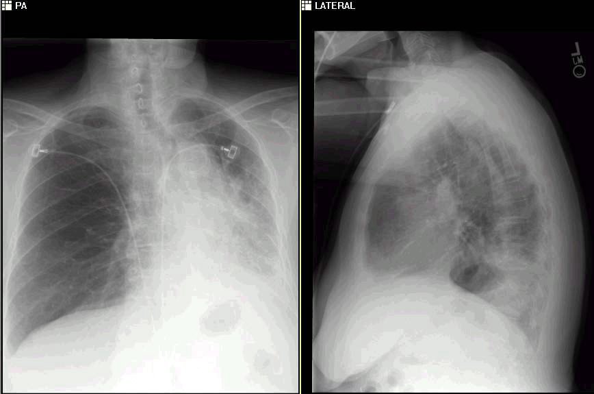
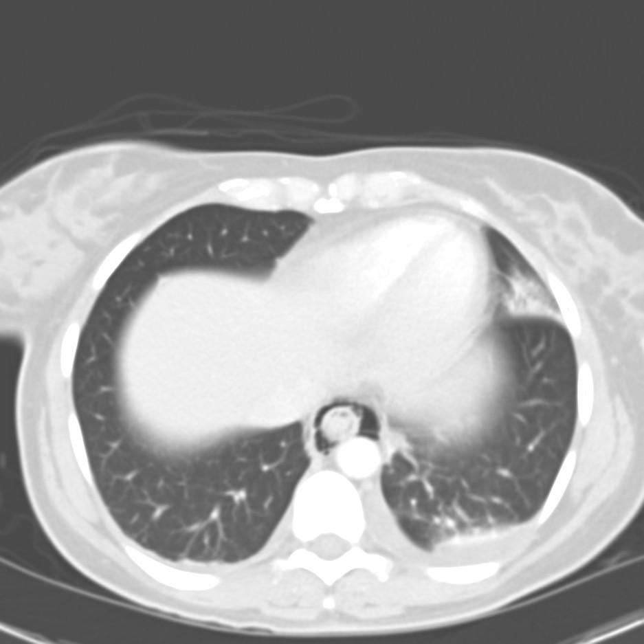
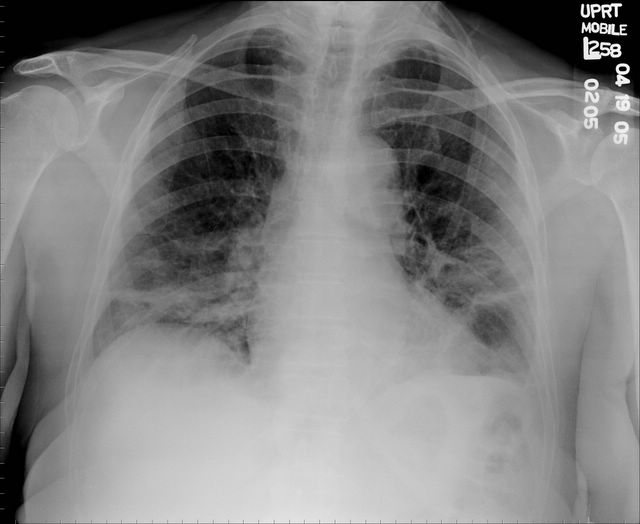
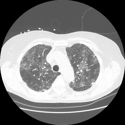
We were consulted on this pleasant 58-year-old white male with known coronary artery disease. He was last admitted 3 months ago with a inferior lateral wall myocardial infarction. He is status post angioplasty. He has a history of chronic obstructive pulmonary disease. He had been doing fairly well andhad gone back to work as an electrician however, over the last five days he developed increasing lethargy, chills and felt feverish with a dry nonproductive cough and worsening dyspnea.
PMHx: Ventricular tachycardia, S/P AICD/PM. CAD, S/P inferior lateral myocardial infarction and previous EF~45%. He is status post stenting of the OM/circumflex. Chronic obstructive pulmonary disease. Dyslipidemia.
Current meds: Lovenox, Albuterol and Atrovent, Levofloxacin, Aspirin and Amiodarone.
FHx: Noncontributory
SHx: Heavy cigarette smoker, he quit 3 months ago. He has been drinking several beers daily. He works as an electrician. He lives alone and his wife died last year.
ROS non-contributory.
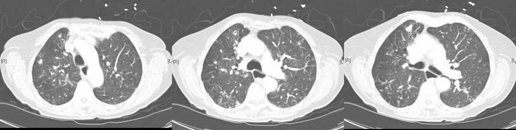
On exam he was tachypneic to 36 with an O2Sat of 90% on BiPAP with 100% FiO2. BP and HR were stable. His lungs had very faint crackles and no wheezing or ronchi.
ABG was: 7.43/CO2: 37/ O2: 58 on 100% FM.
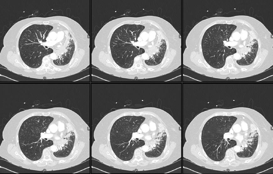
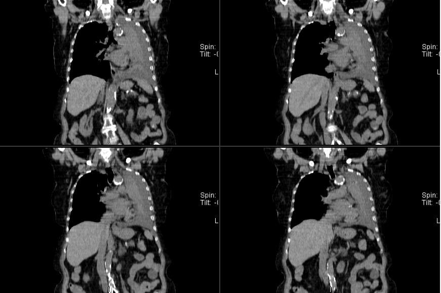
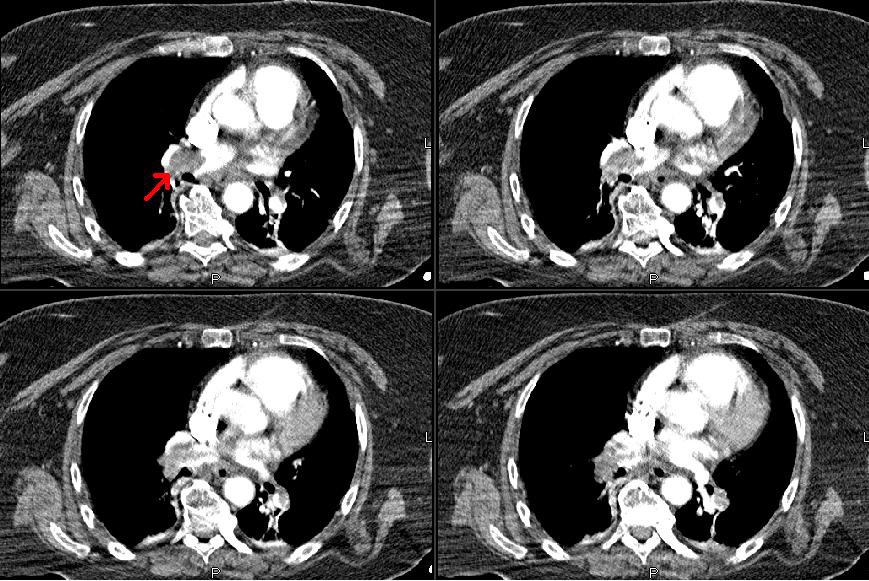
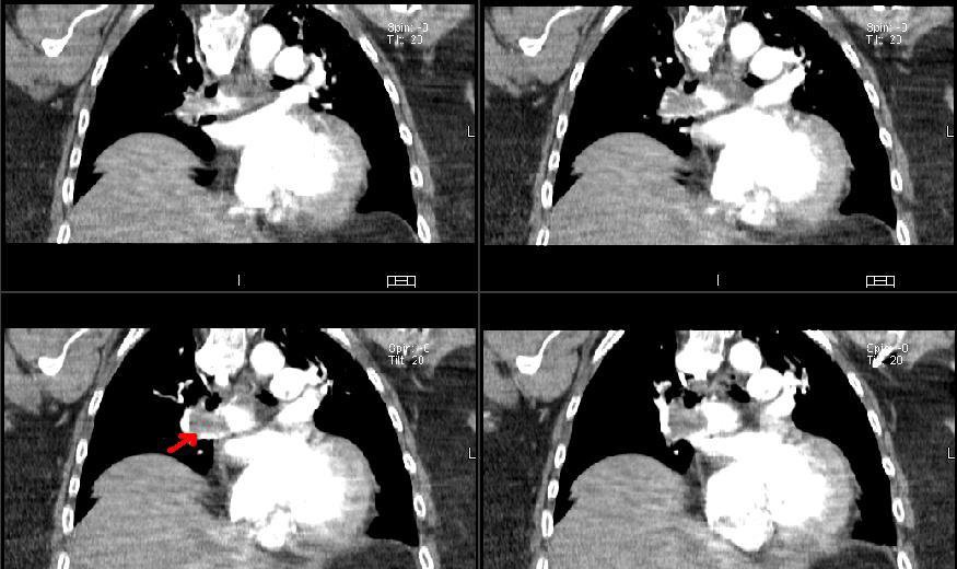
His CxR and CT are posted below.
What would you do next?

Monday, November 28, 2005

ICS in COPD

This is a quick question. How often do you add inhaled steroids in the care of your COPD patients? I am more curious about people's anecdotal and personal opinions rather than data (FEV1< 30%, response to PO steroid trial, etc.).

Thursday, November 24, 2005

Happy Thanksgiving

We will be back soon with more posts.

Tuesday, November 22, 2005

Follow-up to Picture Friday (Janeway lesions)

This is the 65 y/o man with FUO and palmar and plantar lesions. He has now had about 4 weeks of ABTx. His serologies for Coxiella and RMSF (and other Rickettsia) have been negative. His TEE showed some abnormal signal at one of his pacer leads but no vegetations. He remains afebrile, his Janeway lesions and splinter hemorrhages are almost completely gone and his arthralgias resolved. A Bx of one of his lesions was read as embolic infectious vasculitis. We still don't have an organism but with his good clinical response I have called it "culture-negative endovascular infection" and he will have a total of 6 weeks of therapy because of the hardware (PM). Any other ideas?

Monday, November 21, 2005

Consult to "bronch"

We were asked to consult on a patient to "rule out endobronchial lesion via bronch".
A 52-year-old male has an unremarkable past medical history. He has a 100 pack-year histor and is an alcoholic. He noticed a "boil" over his "right collar bone". He thought maybe he was bitten by an insect. Since then has had progressivesymptoms resulting in chest pain and difficulty breathing.
Blood pressure 120/70, heart rate 120, respiratory rate 22, temperature 36.4. 99% on four liters nasal cannula. General appearance: This is a cachectic male. Lungs sounds were significant for good air entry bilaterally with vesicular breath sounds, no rhonchi or wheeze appreciated. Cardiovascular significant for a tachycardia. otherwise unremarkable. b Over the mid sternum at around T3 is a 1 cm round red maculopapular lesion; no pus from the site.

Thursday, November 17, 2005

Cool films!

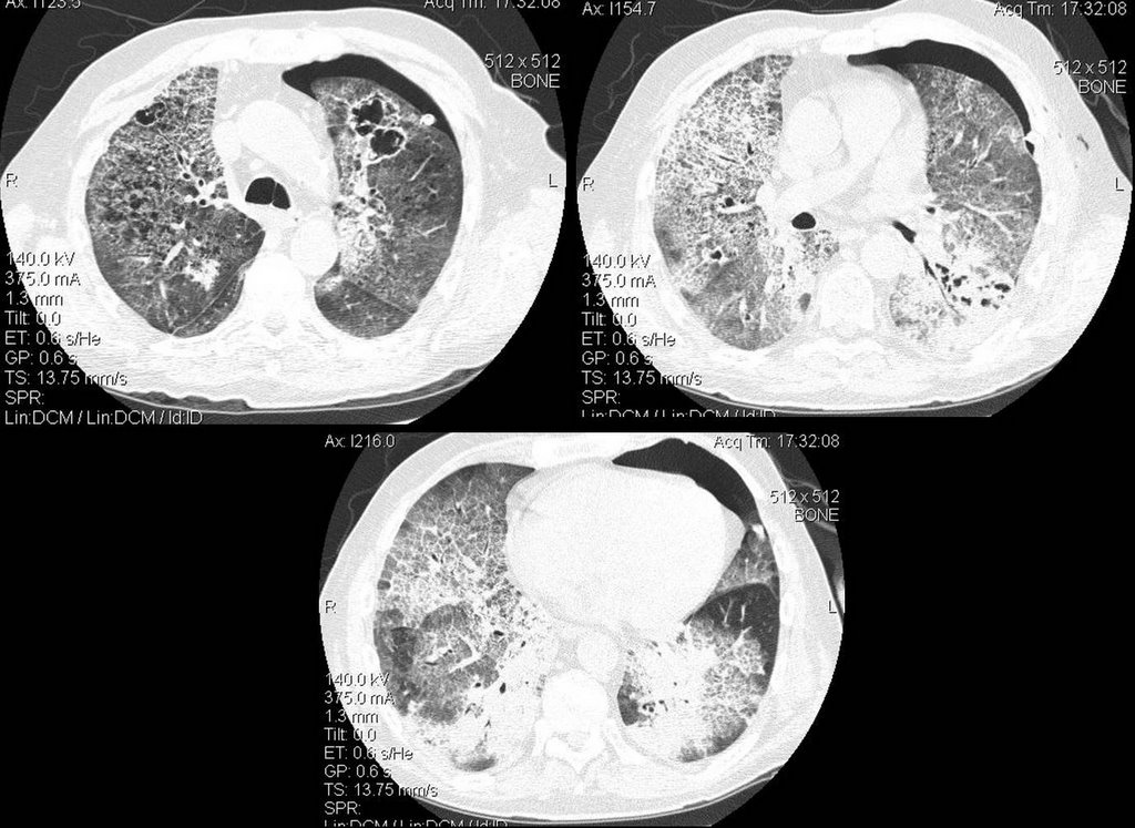
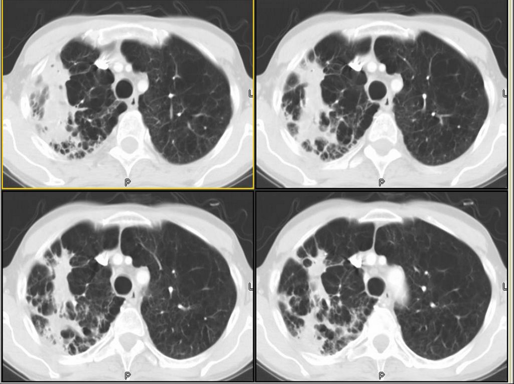
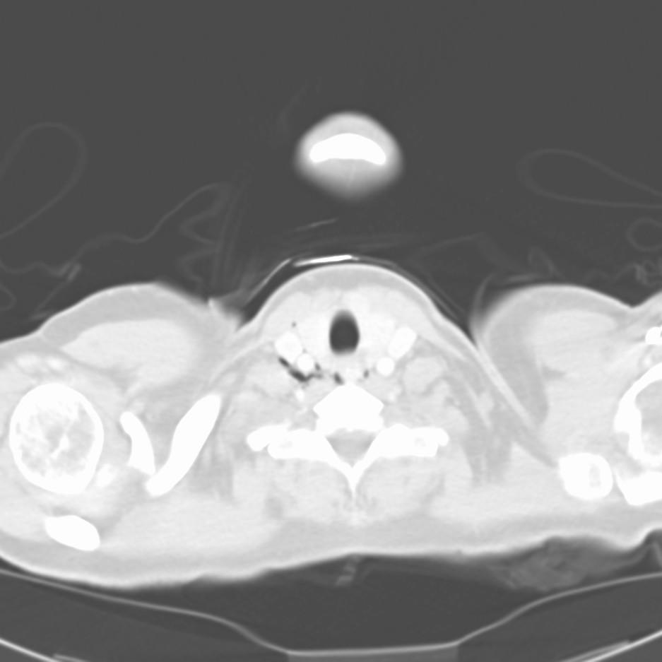
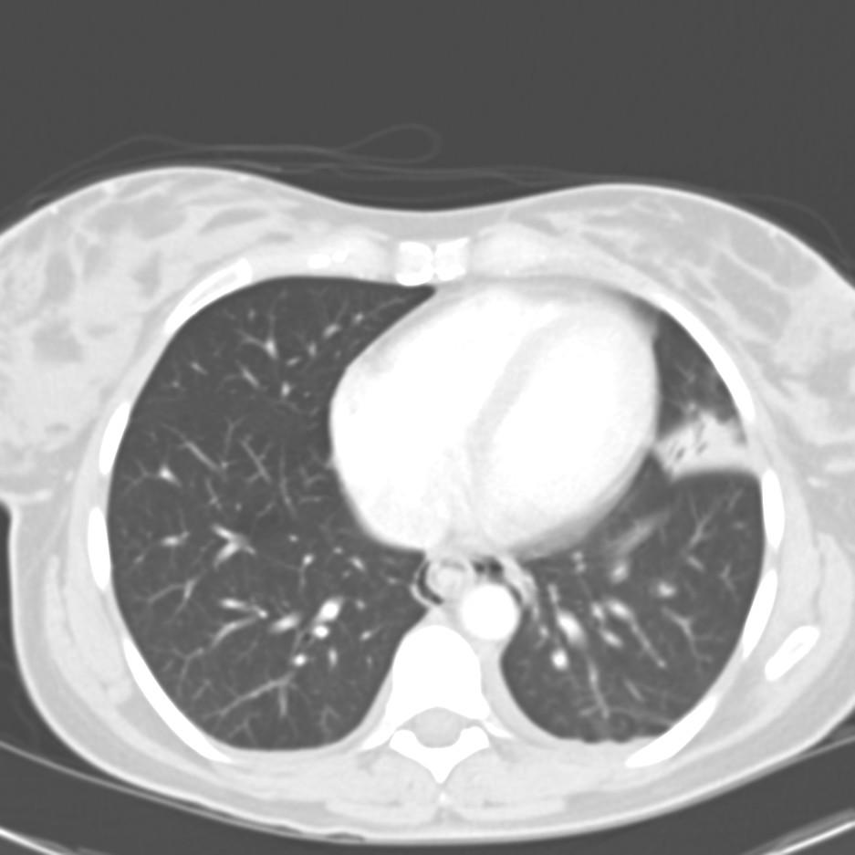
50-something y/o man with no significant past medical history prior to 3-4 months ago, when he developed progressive shortness of breath and required oxygen treatment. His evaluation elsewhere led to a diagnosis of BOOP on a surgical lung biopsy, and he was started on steroids-initially 20mg/day. He failed to improve, and a few weeks later his prednisone was bumped to 40mg/day, and then a few weeks later he had still failed to improve and his dose was increased to 60 mg/day.

A few weeks later, he was admitted to another hospital with continued dyspnea, and transferred to our institution, where he quickly developed respiratory distress. The following CT scan was obtained, and he was transferred to the ICU.


Questions:
1) What intervention led to a rapid improvement in this patients acute respiratory decompensation.

2) What is the the differential diagnosis for the other two findings on this CT scan

3) What steps would you take to further evaluate and treat the patient?

Wednesday, November 16, 2005

Followup to 2 weeks of dyspnea

Mendez and Jeff H both picked up on the salient points. Here is the canned message from Carlos as he hangs out somewhere in Lima:
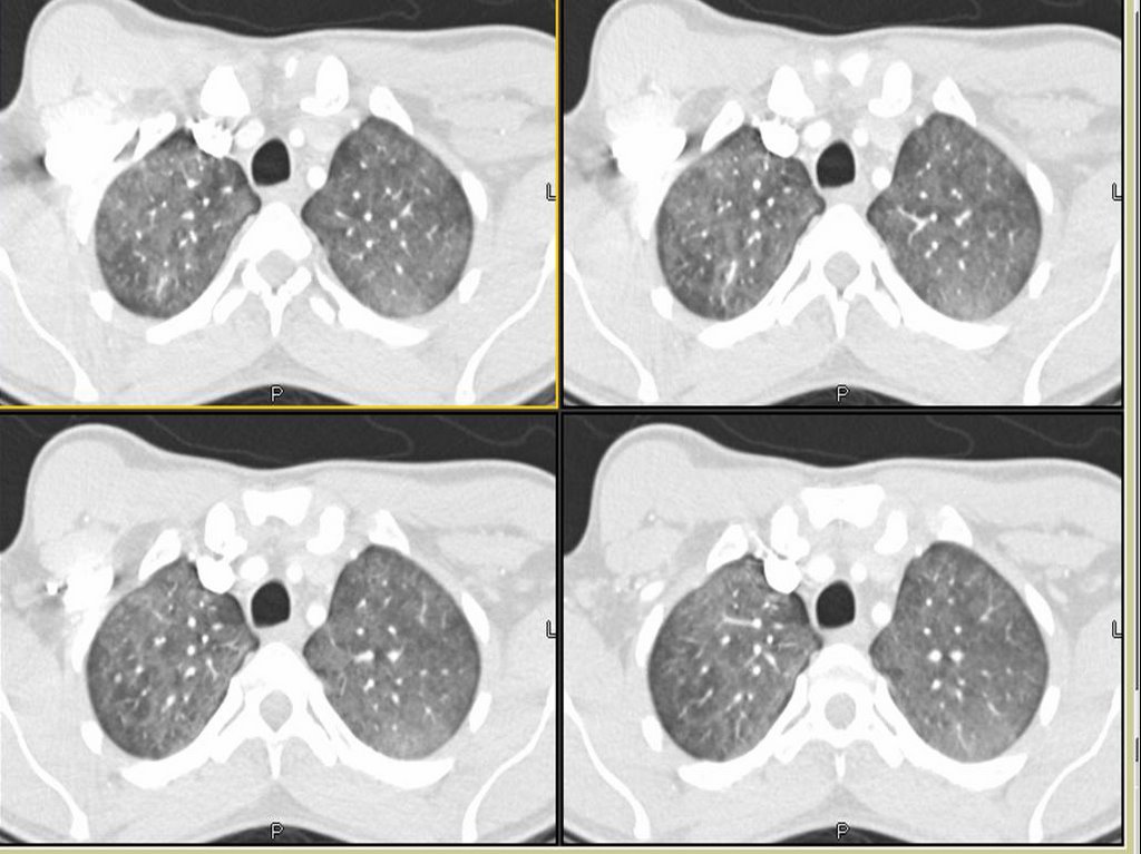
For those who picked up on the "clear lungs" with all the ground-glass we also suspected PCP in this young man with no other Hx of structural lung Dz. He was admitted at night so we started him on IV bactrim and steroids (and a 3 rd gen Cef), ordered a rapid HIV test and planned on doing a bronch. His HIV was positive and then they volunteered his partner was HIV+ and requested a transfer to a University Hospital here in TN. His PCP DNA PCR was positive, his CD4 was around 50 and overall he did very well. Here is his follow-up CxR:

ARDS and pulmonary function

From a PCP in NC:

"Quick question: what PFT parameter is the last to normalize (if it ever does) in patients with ARDS, 1 year out?" The pulmonary boards were yesterday so some of you fellows out there should have no problem with this question.....

Tuesday, November 15, 2005

2 weeks of dyspnea

Carlos' case:
We were consulted on this is a 28 y/o man with no previous PMHx who presented to the ER with progressive worsening DOE over the past 2-3 weeks with some dry cough. Occasional chills but no fevers or night sweats. In the ER he was febrile and hypoxic (7.47/33/58) and was admitted for further care.

PMHx: No lung Dz.

SHx: Occasional ETOH, TOB 1ppd x 10 years, no occupational exposures, no IVDA, lives with same-sex partner.

On exam, AAOx3 in minimal resp discomfort. Clear lungs B/L. S1/S2 RRR and benign abdomen.

CxR:


The ER did a CT-angio (on everybody), which showed no PE but the following changes:


What is your DDx and what would you do next?

Monday, November 14, 2005

Pulmonary embolism and intervention

The cover of the Nov 1st Blue journal (AJRCCM 172(9)) showing a fibrosed thrombus reminded me how controversial treatment of this entity can be. I was interested in what others felt about the following. We all know that no study has demonstrated a change in mortality after thrombolytic therapy for acute massive PE. We also know that some of the beneficial paramters (such as right heart strain or PA pressure) are no different than just IV heparin after about 7 days. Nonetheless, does anyone feel that the lack of a mortality benefit may be as a result of the studies being underpowered? If this is the case, perhaps thrombolytics should be used as default pending a larger study (for massive PE without obvious bleeding risks). On the other hand, if the concensus is that nothing is proven with regards to mortality and thus thrombolytics are NOT indicated by default until that larger study comes along, should we ever administer it?
Now it gets even more sketchy when we consider surgical embolectomy. When do you consider this intervention? Some have argued that it absolutely has no place. Others will ship everyone with a b/l saddle embolus out to the Mayo for embolectomy.
I think there is not a lot of convincing data out there for anything other than good old fashioned anticoagulation and IV fluids (and some prayer).
What do you all do when you get the call about a patient with acute massive P.E.?

Sunday, November 13, 2005

When to Treat MAI

80 yo female with PVOD s/p TIA many years ago presented with multiple pulm nodules about 2 years ago to her PCP. The radiologist was worried about TB at the time, so she was sent to the county health clinic. The CT also showed evidence of broniectasis at that time. Sputum cultures revealed MAI.

She went to an ID doc, who convinced her the treatment is worse than the disease. So, she opted for no treatment.

She is pretty much asymptomatic since 1/04 (the time of her original CT), but she presented to the ER with hemoptysis (about 5 tbsp, bloody sputum with clots). She was watched in observation overnight and it did not recur.

Her repeat CT scan (to rule out PE, of course), showed stable nodules. There were more of them, but the ones visualized 2 years ago are definitively stable.


Would you offer a bronch, or is the CT with bronchiectasis enough to convince you of the diagnosis (she has never before been endoscopically evaluated)?

Would you treat the MAI in this 80 yo, relatively asymptomatic individual?

Tuesday, November 08, 2005

COPD and weight loss.

From Carlos, who is on vacation:
This is a 60 y/o man with a 50 p/y Hx of smoking sent to us because of worsening DOE, cough productive of scant amounts of thick sputum and weight loss. No hemoptysis, no CP.

PMHx: Mild HTN.

SHx: 50 p/y TOB. Quit in 2003.

On exam, thin male in NAD. Distant BS B/L with rare R upper crackles. S1/S2 RRR, benign abdomen.

His Spiro revealed an FEV1 of 1.1 L (27% of his predicted) and he had the following CxR and CT scan findings:


update(after 1st 2 comments):
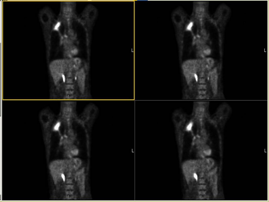
He was started on aggressive bronchodilator therapy, given ABTx and steroids and returned with symptomatic improvement. His FEV1 increased to 2.17L (53%) and he felt better. However, he still had some weight loss (6 lbs.) and a persistent infiltrate after 4 weeks. I did a bronch which revealed only chronic bronchitis and no other Dx.

A PET-CT was done and it revealed marked FDG uptake to the RUL (average SVU of 13 with max SVU of 21) and nowhere else. What would you do next?

Monday, November 07, 2005

18 year old with hemoptysis

18-year-old male referred for hemoptysis. He reports several episodes of pneumonia in last several years. His mother believes that these have all been on the "right chest." The most recent episode of pneumonia had occurred during the last football season, when he developed cough and hemoptysis. By report, he had an abnormal chest x-ray at that time. He was treated with antibiotics and reports that the symptoms resolved. He again had hemoptysis in a few months later. A PPD was negative. No images of a chest CT, but there was right middle lobe bronchiectasis.

Pmhx Status post hernia repair, rhinoplasty, right elbow surgery. In addition, the patient has a history of a racing heart and has been worked up for possible supraventricular tachycardia. In addition, the patient had a mole punch biopsied off his left back inferior to the scapula and does not know what the pathology for this showed.

SH: no alcohol, tobacco, or drug use

Physical Exam: His P is 49. BP is 108/57. His RR is 16, and his WT is 79 kg. In appearance, he is a well-developed, well- nourished young man in no distress. HEENT: Lymph node examination was no supraclavicular adenopathy. Pulmonary: clear to auscultation with both quiet and forced expiratory maneuvers from base to apex bilaterally. He is resonant to percussion throughout. Cardiac examination: Regular rhythm. S1 and S2 are of normal quality without murmur, gallop, or rub. He had no pedal edema, and he had 2+ radial pulses.

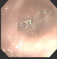
A bronch showd the following in the RML:
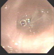
My collegue proceeded to evaluate further into the bronchus where they identified copious mucous and a foreign body. The foreign body was extracted with biopsy forceps. Under direct visualization it appeared to be a piece of organic green matter that resembled a branch tip of a pine tree.
Final diagnosis:
LUNG, RIGHT MIDDLE LOBE, BIOPSY:
1. BENIGN MARKEDLY INFLAMED BRONCHIAL MUCOSA AND FRAGMENTS OF
GRANULATION TISSUE.
2. FOREIGN MATERIAL CONSISTENT WITH VEGETABLE PARTICLE.
B. RIGHT LOBE, BIOPSY: FOREIGN BODY.

The patient breathed in a pine needle....

Friday, November 04, 2005

Approach to this lesion

A physician wants some feedback regarding how to approach this lesion. This is an abdominal CT. A chest CT has not yet been done. The quesion regarding approach: get a PET? Go right to CT-guided biopsy? Get a full CT of chest first in case soemthign shows up that would make a bronch more helpful?
He's a patient with COPD and heavy smoking history. Abd CT was obtained b/c of some nausea.

Thursday, November 03, 2005

Check out our ongoing cases!

Just a reminder to check on some of the ongoing cases.
DKeena had a great case on Enbrel-associated infiltrates in need of comments.
I have posted follow-ups to the drop in ETCO2 case (check out the CT) and the abnormal CxR as well as the presumed IE.
And we are looking forward to more info on Jennings' case with pancytopenia and abnormal lungs.

Update on "update on picture Friday"

This is the 65 y/o man with FUO and palmar and plantar lesions posted on Picture Friday. There have been a few very good new comments on the original post. Dr Jon wrote:
Rickettsial infection: what about R. prowazekii? More common in elderly, and consistent with petechiae, but not common in US. Has he ever had typhus? There is Brill-Zinsser Disease (has he ever traveled outside th country?)Also about the endocarditis, maybe C. psittaci (hard/impossible to culture), does he have any birds?
He did not have birds and no exposures to suggest typhus. No recent travels. His serologies for Q fever, RMSF, Rickettsia prowazekii and Ehrlichia came back today and were all negative. He has remained afebrile and his Janeway lesions are resolving. A skin punch biopsy was interpreted as infectious embolic vasculitis though no organisms were seen. All Cxs remain negative. A TEE showed no veggies but there were some echo changes at the PM atrial lead site concerning for a small endocardial abcess. His CRP is down from 30 to 6 but now his Creatinine has crept up from <1.5>3%.
So we are still treating him for Cx- IE. Any other ideas?

Follow-up to abnormal CxR - part II

This is the woman in her 70s with an abnormal CxR. After the thoracentesis she had more L lung collapse: by the time of the bronch and the f/up PET she had complete L lung collapse (making her at least a IIIA). Her past showed uptake only in the peri-hilar mass itself. Our surgeons and a CT surgeon from a large University Center nearby felt that though she was a IIIA, her lesion was very proximal (less than 2 cm to the carina) and she would be a poor candidate for a pneumonectomy so she is receivin Chemo and xRt. She will follow-up with us and may benefit from APC or stenting to the lesion to improve drainage/clearance of the collapsed lung.

Wednesday, November 02, 2005

Unbound Medicine

We came across this jewel of a site from Mexico that focuses on medical education. As such there are some interesting cases posted there.

Follow-up to abnormal CxR

This is the woman in her 70s with an abnormal CxR. Everyone picked up on the loss of volume and nobody was "fooled" by the negative CT last year (it was really unremarkable). She had a CT scan seen below:

Most of her L lung was collapsed with a smallish effusion. Her effusion was exudative with negative cytology. I did a bronch and she had a NSCLCa occluding her L mainstem bronchus. Her Spiro (with her L lung collapsed) revealed an FEV1~ 800cc.
What would you suggest as the next step?

Follow-up to drop in ETCO2

Good comments on this case. A drop in ETCO2 suggests a massive mismatch or loss of pulmonary vascular perfusion. Cardiac arrest would do that but I did not describe full arrest. DKeena mentioned esophageal intubation. It can be tricky especially in emergencies when a lot or air is bagged into the stomach and an initially +ETCO2 drops as the tube stays in the esophagus. Massive PE will cause a decrease in CO2-rich blood flow to the lungs. Since her ETT was properly placed and her EKG showed no ischemic changes (just the LAFB) and she is a sedentary (having back surgery, remmember?) I was concerned she had had a PE and requested and emergent CT. Check it out below:


Would you thrombolyze her?
What would you do next?

Tuesday, November 01, 2005

Quick follow-up to Asthma (with Strongyloides)

This is the 20 y/o woman with bronchospasm and cough after her missionary trips. She has finished her course of Albendazole and has been on Asmanex (mometasone). She is now assymptomatic, her lungs are completely clear and her FEV1 increased from 2.29L(76%) to 3.30L(110%) and her ratio increased from 80% to 87%. We are weaning her Asmanex off and her repeat stool O&P were negative.

Pancytopenia, abnormal cxr

I hope to have a lively debate because there is some controversy about the following case:
34 y/o AA woman with pancytopenia 10 years ago. Asymptomatic, lost to f/u because of insurance. Refused BM biopsy. 5 years ago gradual onset of dyspnea. May of this year presented with SOB and admitted. (Pulmonary consult called at that time). On exam she had some JVD, clear lungs, loud O2, hepatosplenomegaly, ascites and 2+ pitting edema. Echo showed PAP of 48 with dilated RV and an LV with an EF of 65%. Labs: WBS 2.2 with 82% N, Hgb 9, plts 104. ACE was 207.
CXR and CT:

Bronch BAL not sent, therefore cell count and micro of the fluid unknown. A TBBX showed of the RNL showed a single poorly formed granuloma. The micro on the biopsy *was* sent and was negative. Endobronchial biopsy showed active chronic inflammation.
Urinary histo Ag never sent. Serum Ab's against fungus also not sent.

Questions:
1. Would you consider the above info sufficient stop at the diagnosis of sarcoid?
2. Would you be satisfied with no infectious cause based on negative Tbbx micro, but BAL not sent?
3. Do you feel that a cell count with CD4 and CD8 would have been helpful to you?
4. (corollary to 1): Is there anything else on your differential list besides sarcoid? The person presenting this case to me has no other diagnoses on his list.
5. Does the history of 10 years of pancytopenia rule out histoplasmosis or other fungus?

Monday, October 31, 2005

Abnormal CxR

We were asked to see this 74 y/o woman for dyspnea and atypical chest pain. She has a long TOB Hx (>50 p/y) and continues to smoke. Last year she had a "lump" in her neck (it was benign) and had an unremarkable neck and chest CT scan. For the past 2 weeks she has had worsening dyspnea and a cough productive of blood-streaked clear sputum.
No othe significant PMHx or exposures.
Her exam reveals decreased BS on the L with faint crackles and her CxR is seen below:

What would you do next?

Drop in ETCO2

This was an interesting consult: 75 y/o woman with depression (lost her husband a month ago) and chronic back pain came in for an elective laminectomy. She received rapid induction with Propofol and succinylcholine and was intubated rapidly. She then had sudden bradycardia and hypotension with hypoxemia and her continuous end-tidal CO2 indicator dropped precipitously.
They cancelled the surgery and gave her dopamine and fluids. She had a rebound tachycardia with dopa, was given neosyn. and cardiology and I were consulted.
What other info would you want?

Update on Picture Friday case

This is the 65 y/o man with FUO and palmar and plantar lesions.
Since the post his temperature curve has come down nicely on ceftriaxone and doxy. He still has the skin lesions and blood Cxs (after 7 days incubation) are still negative. He is having a TEE today and RMSF and Coxiella serologies are pendng. His RF was negative but he had a +ANA with +Anti-RNP (Anti-DS-DNA, Anti SSA and SSB and other SLE markers are negative) and his CRP has come down from 30s to 20. Any other ideas?

Friday, October 28, 2005

Pulmonary infiltrates from etanercept

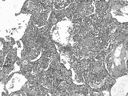
So to make a complicated story more complicated- when I spoke to our pathologist about the biopsy results he said "I never seen anything quite like this in my career." A prominent lung pathologist from another institution was in agreement.

I've posted their description of the findings below:

Lung, right, middle lobe, wedge biopsy:
Chronic interstitial pneumonia and patchy interstitial fibrosis; chronic active bronchiolitis with focal proliferative-type bronchiolitis obliterans and organizing pneumonia pattern; focally necrotic granulomatous inflammation.

A constellation of pathologic findings are present in this case. The alveolar parenchyma manifests chronic inflammatory cell infiltrates as well as patchy
fibrosis. Lymphoid aggregates are present, and many of the interstitial cells are plasma cells. Superimposed on this picture are numerous granulomas, some of which show focal necrosis and acute inflammation. A few of the granulomas appear bronchiolocentric. Rare bronchioles contain intraluminal foci of granulation tissue. Special stains are negative acid-fast bacilli and fungi.
The background interstitial inflammation and fibrosis may be related to the patient's rheumatoid arthritis.

A recent publication (Modern Pathology 2005 18:651) describes a series of patients who developed persistent pulmonary infiltrates and granulomatous inflammation associated with etanercept use. The histopathologic changes noted in those patients was similar to what my patient had; here is a pic:

In that series, all of the patients improved with a combination of immunosuppressive therapy and discontinuing etanercept.

This patient has clinically stabilized but has been slow to improve.

Posted by Jennings for Dan.

Thursday, October 27, 2005

Picture Friday

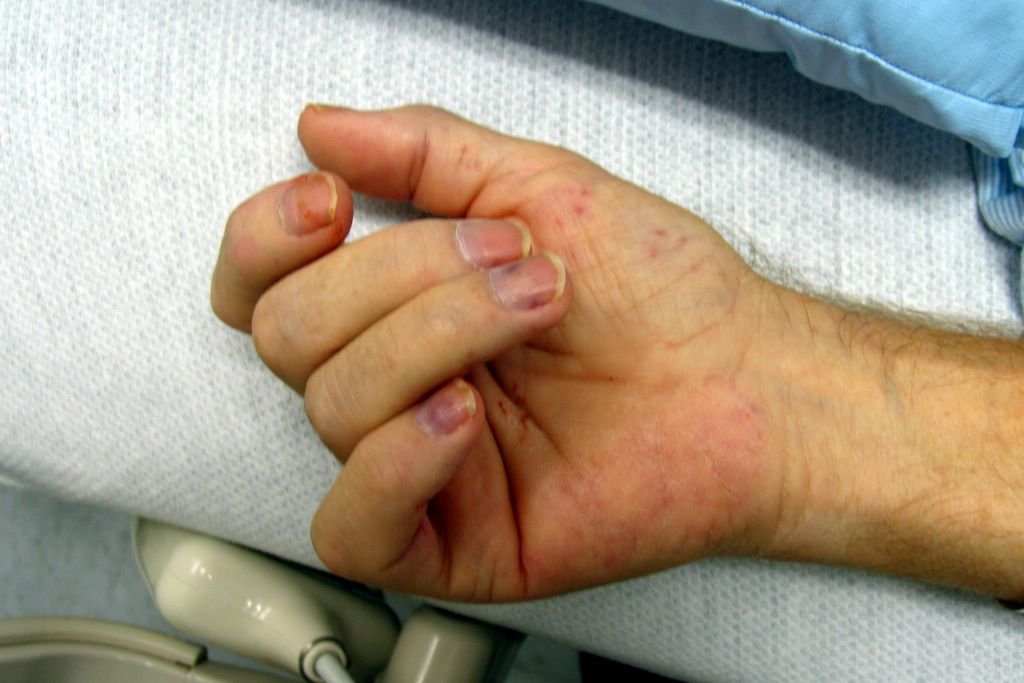
We were asked to see this patient with FUO. This is a 65 y/o man with CHF (EF~25-30%) with 6 weeks of daily fevers, now with arthralgias. He has been admitted twice with multiple negative Cxs and no diagnosis yet.
He has clear lungs, no arthritis (just arthralgias) and the following findings on hands and feet:

Wednesday, October 26, 2005

Radiograph of the day

87 year old man with no PMH presented to his PCP with mild SOB. No cough. No constitutional symptoms. He is a non-smoker.

The CXR is shown:

Answer below with discussion.

Tuesday, October 25, 2005

Fever and chest pain post-EGD

This is my Wednesday case; feel free to email the posting schedule, however.

35 yo female post-partum x 7 weeks (healthy newborn girl; no complications with pregnancy or with birth) with no PMH presented with the sensation of food getting caught in her throat after eating a large steak meal.
EGD was performed with foodstuff visualized in the proximal esophagus. It was pushed successfully into the stomach.
Post-procedure, the patient had onset of fever (38.5) on one occassion and mild chest pain that resolved without intervention.
Her WBC count was normal and did not change throughout her hospital course.
A CT chest was obtained to evaluate.


How would you proceed?

Answer below with discussion.

Dyspnea

After "Nodule Week" maybe we should have "Dyspnea w/up week"...
Anyway, I see a lot of older patients with no obvious structural pulmonary or cardiac abnormalities and just want to compare notes.
To ilustrate it, yesterday I saw this 60 y/o woman with "episodic DOE". She is fairly healthy, exercises regularly on a treadmill and was sent by the cardiologists after a normal radionuclide stress test (normal EF too). She notices dyspnea with chest tightness when she tries and exerts herself "faster than usual" like racing up the stairs. Her daughter got married a few weeks ago and everytime she went up to get something she would get dyspneic. No F/C/NS/cough.
No TOB. Occ social ETOH.
On a PPI, Calcium and prn Tylenol.
Normal exam (normal BMI!).
Normal PFTs (FEV1 and ratio, TLC, no air trapping and DLCO~101%).
She had had a normal chest CT and we did a Mathacholine test which was negative (~10% change in FEV1 after 25mg).
What would you do next in this case? Do you see a lot of this less obvious cases of DOE?

Sunday, October 23, 2005

Asthma mystery

From Doug:

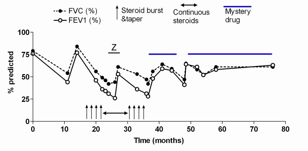
This is not so much a diagnostic dilemma, as a therapeutic curiosity. Think of it as a riddle. This is a very nice lady whom I cared for about 5 years before she was lost to follow up because of an insurance change. She initially came to my clinic after being followed by another colleague who left the University after finishing training. This lady had very severe asthma, with persistent symptoms, and frequent exacerbations that required steroid burst & tapers. She was also quite obese and as a result had some restrictive physiology that was not accompanied by any evidence for interstitial disease on full PFTs and HRCT scans done on multiple occasions throughout the years. After a while I ran completely out of options for her. She was compliant on 6 puffs of Flovent bid, Serevent, Accolate (this was before Singulair), et., and was still ending up in an emergency room every 4-6 weeks.

I started her on a drug that we shall henceforth refer to as "Mystery Drug". I had a biologic rationale for it, but NO DATA whatsoever, other than I knew it to be acceptably safe. I will see if anyone can guess what I started her an and why, then post some follow up.

Thursday, October 20, 2005

Cough with bronchiectasis

A 44 y/o woman was referred to me for possible atypical mycobacterium. Several years ago, she was diagnosed with MAC and treated for 6 months with Rifampin, Ethambutol, and Biaxin. Her therapy was stopped because of optic neuritis. Over the following several years, she remained stable, but in poor general health. Recently, she has developed recurrent sinusitis, and has had several complicated sinus surgeries. She was referred because of persistent cough.

The remainder of her history is unremarkable, with the exception that she continues smoking regularly (1ppd x 20+ years).

On exam, she is thin, but in no distress. Her general exam is unremarkable, although she has some scattered wheezes and rales bilaterally.

Her pulmonary function shows mild obstruction, with normal gas exchange.

A high resolution CT scan shows mild bilateral bronchiectasis, primarily in the upper lobes, middle lobe, and lingula.

How would you approach this patient? I'll post some follow-up as we go...
Follow-up to pulmonary nodule

This has been "Pulmonary Nodule Week on Pulmonary Roundtable"!
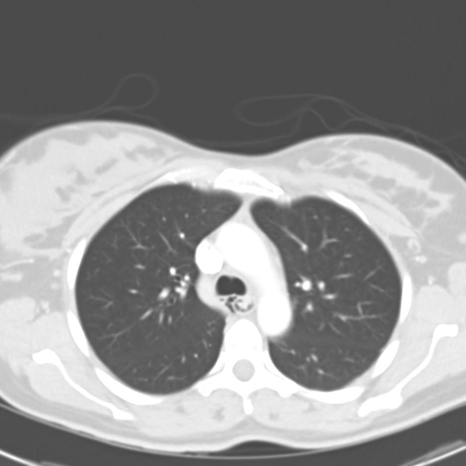
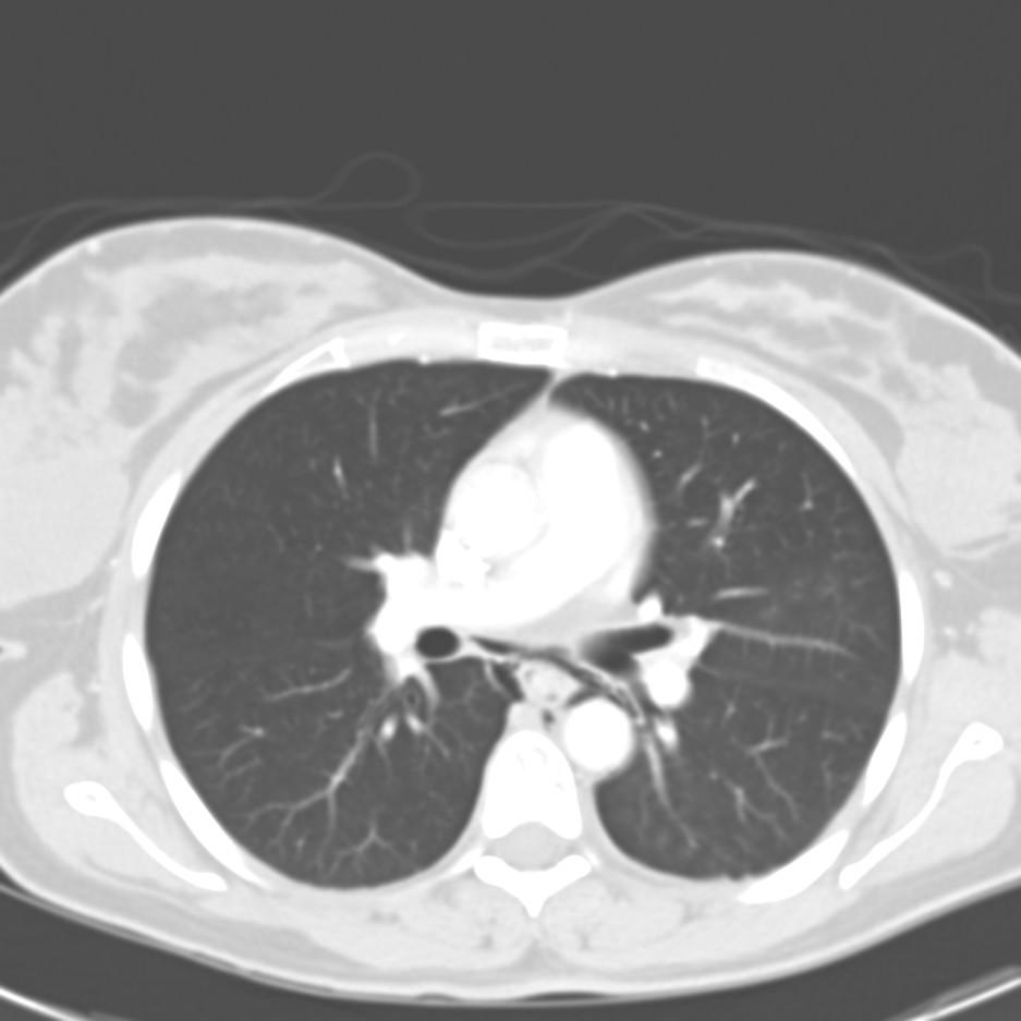
This is a follow-up on the 65 y/o former smoker with poor PFTs and the suspicious lesion. After the poor response to bronchodilators we did perform a PET: it revealed significant uptake by the nodule and no other abnormal areas. We discussed it with our CT surgeons who felt very hesitant about a lobectomy. Instead they did a mini-thoracotomy wedge resection of the area. It revealed a 2x2x1.8 cm poorly differentiated squamous cell-Ca. We have consulted hem-onc and since this was a wedge and not a full lobectomy they have recommended ChemoTx/xRt. We don't do RF ablation here so I felt the wedge with follow-up Tx was as close to it as we would get. She has done very well post-op and should be home soon (surgery was 2 days ago) with hem-onc f/up.
Wednesday, October 19, 2005

More pulmonary nodules

This 49 y/o woman was referred to me with an anormal CT scan. She has a 30 pack/year Hx and has quit smoking when told about the CT. She has no symptoms (she had had a "screening" CxR which prompted the CT), her exam is quite unremarkable and she has normal PFTs (FEV1 and FVC are >85% though the ratio is a bit low).
Her CT showed multiple non-calcified nodules (some a depicted below) with no adenopathy.
What would you do next?

Tuesday, October 18, 2005

Answer to Mike's Pulmonary nodules

Meanwhile, Mike L has posted the f/up and diagnosis on his pulmonary nodules case.
Check it out and leave your comments.

Pulmonary nodule

This is a 65 y/o former smoker who was sent to us for evaluation of a pulmonary nodule. She has COPD, has baseline DOE and while having an AECB had a CxR (outside films). The CxR revealed a nodule which is better characterized on the CT below.

She has had no F/C/NS, no hemoptysis.
Her exam was fairly benign. Her FEV1 however, was only 700cc on Combivent?
What would you do next?

Followup to shortness of breath

Doug picked up on the subtle clue of chronic UTI's on past history.
Here is the xray

On nitrofurantoin


1 month after stopping the nitrofurantoin:
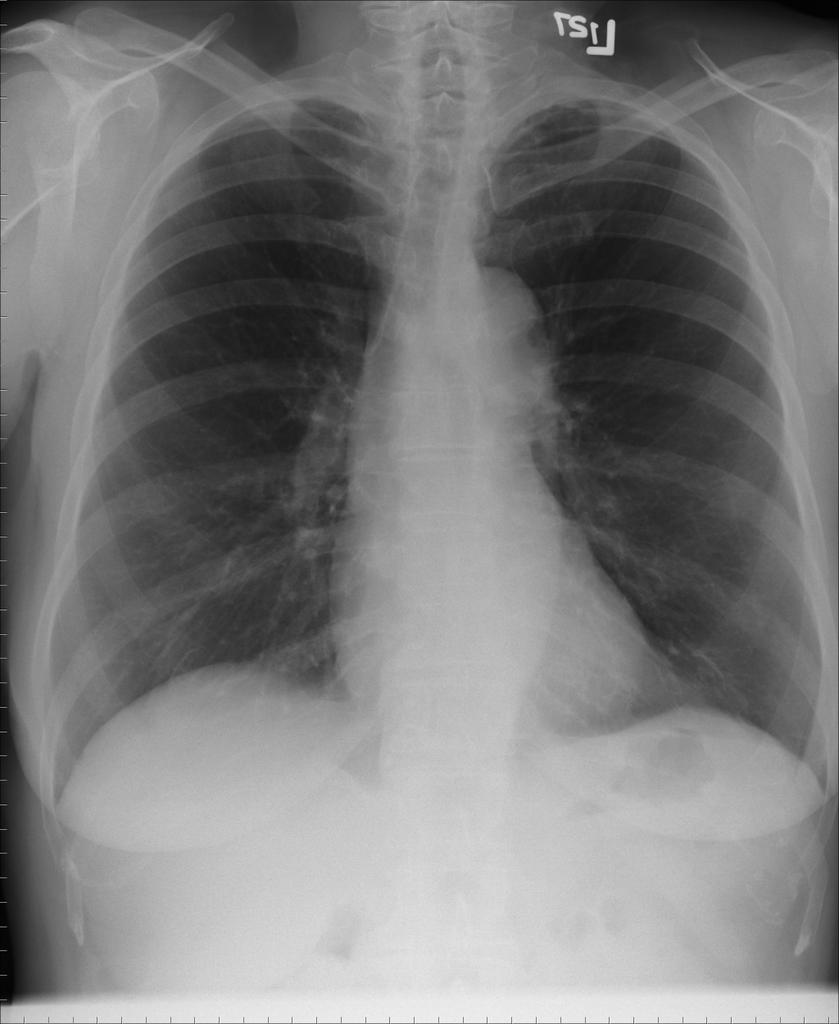
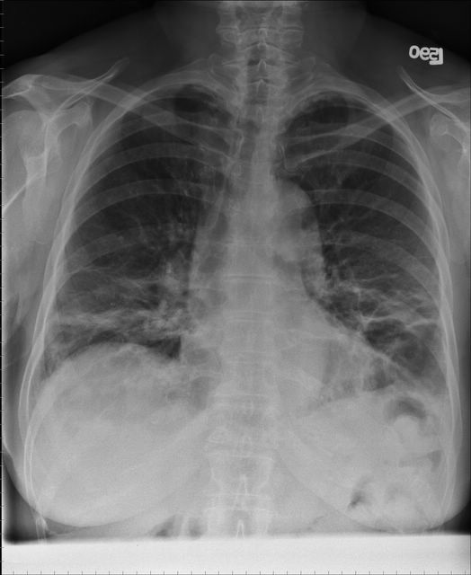
Her shortness of breath resolved and the DLCO (which was 56%) became 86%.

Monday, October 17, 2005

Shortness of breath

74 year-old woman who reports gradual onset of dyspnea on exertion over the past year. No chest pain, fevers chills or night sweats. She has a minor non-productive cough.
ROS: Some minor arthralgias, probably OA; o/w unremarkable.

PmHx: chronic UTI's followed by urology otherwise negative.

SH: Lifelong nonsmoker. Occupation was secretary.
FH: 2 daughters with fibromyalgia.
Travel Hx: Thailand in the past year.

On exam initially the lungs were clear to auscultation, but subsequent exams over the next year revelaed bilateral crackles, as can be imagined based on xrays shown below.

So over the next year she became more and more short of breath with the following progression on chest xray.
First CXR
CXR a few months later

CXR more months later with worsening dyspnea.

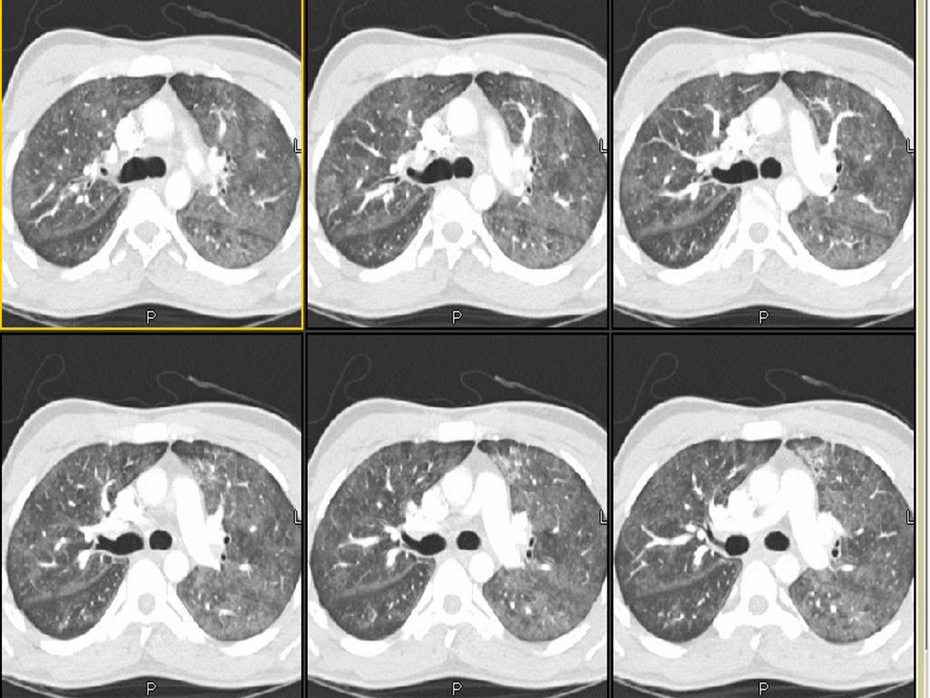
Here is a few CT slices around the time of the second chest xray (above):


ct up

ct mid

ctlow
What additional information would you like to know?

RF and ANA were low in the past.